Pal Cadaver Axial Skeleton Skull Lab Practical Question 4
Onlines
Mar 17, 2025 · 6 min read
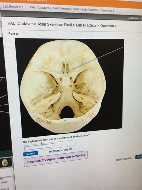
Table of Contents
Pal Cadaver Axial Skeleton Skull Lab Practical Question 4: A Comprehensive Guide
This guide delves deep into a common lab practical question: identifying structures within the palatine bone and surrounding areas of the skull using a cadaver specimen. We'll cover the palatine bone's anatomy, its relationship with other cranial bones, potential lab scenarios, and strategies for accurate identification. This detailed approach aims to equip students with the knowledge and confidence to excel in their practical examinations.
Understanding the Palatine Bone: Anatomy and Function
The palatine bone, a paired bone located within the facial skeleton, plays a crucial role in forming the hard palate, nasal cavity floor, and parts of the orbits. Its complex shape and articulation with multiple bones make it a key focus in anatomical studies. Let's break down its key features:
Key Anatomical Structures of the Palatine Bone:
-
Horizontal Plate: This broad, flat portion forms the posterior third of the hard palate, contributing significantly to the roof of the mouth. Its roughened superior surface articulates with the maxillary bone, while the inferior surface is smooth and contributes to the oral cavity.
-
Perpendicular Plate: This thinner, rectangular plate extends superiorly from the horizontal plate to contribute to the lateral nasal wall and the floor of the nasal cavity. Its medial border articulates with the vomer and the opposite palatine bone, contributing to the formation of the nasal septum.
-
Greater Palatine Foramen: Located on the posterior border of the horizontal plate, this foramen transmits the greater palatine nerve and vessels, providing sensory and vascular supply to the palate.
-
Lesser Palatine Foramen: Found posterolateral to the greater palatine foramen, this smaller foramen allows passage of lesser palatine nerves and vessels, also supplying the palate.
-
Sphenopalatine Foramen: While not directly part of the palatine bone, its location (posterior to the perpendicular plate where the sphenoid, palatine, and maxillary bones meet) makes it crucial to understand its relation to the palatine bone during practical exams. It allows the passage of nerves and vessels from the pterygopalatine fossa.
Articulations of the Palatine Bone: A Key to Identification
The palatine bone's unique shape and its articulations with multiple bones are crucial for its accurate identification during a practical examination. Here's a detailed look at its key connections:
-
Maxillary Bone: The horizontal plate articulates with the palatine process of the maxilla, forming the majority of the hard palate. This articulation is a key landmark to confirm palatine bone positioning. Look for the relatively smooth palatal surfaces of both bones which seamlessly connect.
-
Vomer: The perpendicular plate of the palatine bone articulates with the vomer, a midline bone of the nasal septum. This helps in orienting the palatine bone within the nasal cavity. Pay attention to the subtle interlocking edges of these two bones.
-
Sphenoid Bone: The perpendicular plate also articulates posteriorly with the sphenoid bone. This articulation contributes to the posterior part of the nasal cavity floor and is vital for understanding the overall craniofacial architecture. This connection is often more complex and requires careful observation.
-
Ethmoid Bone (Indirect): While the palatine bone doesn't directly articulate with the ethmoid, their close proximity and involvement in the nasal cavity make understanding their spatial relationships essential.
-
Zygomatic Bone (Indirect): Similarly, the relationship of the palatine bone to the zygomatic bone, even indirectly, is significant when visualizing the overall architecture of the mid-face.
Lab Practical Scenarios and Identification Strategies
Practical exams often involve identifying specific structures on a cadaveric skull. Here are potential scenarios and effective strategies:
Scenario 1: Identifying the Palatine Bone within the Maxilla:
- Strategy: Begin by locating the maxillary bones, then carefully trace the palatine processes of the maxilla. The posterior border of the maxillary palatine process will meet the anterior border of the palatine bone. Feel for the smooth junction to confirm articulation. Look for the Greater Palatine Foramen as a landmark.
Scenario 2: Locating the Greater and Lesser Palatine Foramina:
- Strategy: After identifying the palatine bone's horizontal plate, locate the posterior border. The greater palatine foramen is typically more prominent and sits slightly anterior to the lesser palatine foramen. Use a probe to gently explore the foramina and verify their connection to the pterygopalatine fossa.
Scenario 3: Differentiating the Palatine Bone from Adjacent Bones:
- Strategy: The palatine bone's unique shape and the precise articulations are crucial differentiators. Note the different textures and contours of the bone's surfaces. The maxillary bone has a more substantial palatine process with a rougher texture compared to the smoother surface of the palatine bone.
Scenario 4: Identifying the Palatine Bone in the Nasal Cavity:
- Strategy: Look for the perpendicular plates of the palatine bones that contribute to the formation of the nasal cavity's lateral walls. Their articulation with the vomer is crucial for orientation. Carefully probe the region to identify the various bone edges and their relationships.
Potential Challenges and Tips for Success
-
Bone Fragility: Cadaveric bones can be fragile; handle them with care to avoid damage.
-
Overlapping Structures: The complexity of the craniofacial anatomy means bones often overlap. Carefully separate and expose the structures of interest.
-
Individual Variation: There's natural anatomical variation. Don't panic if your specimen slightly differs from textbook illustrations. Focus on the general principles and relationships.
-
Using Tools: Practice using dissecting probes and other tools to carefully explore the foramina and articulations. Learn to use the right amount of force to avoid damage to the bones.
-
Pre-lab Study: A thorough understanding of the bone's anatomy, location, and articulations is critical. Use anatomical models, videos, and textbooks to build a strong foundation.
-
Collaboration: Study with classmates to learn from diverse perspectives. Discuss your findings and approach.
Beyond the Lab Practical: Clinical Significance
Understanding the palatine bone is not just about passing an exam; it has significant clinical relevance. Its involvement in cleft palate development, dental implants, maxillary sinus surgery, and other procedures underscores its importance. The knowledge gained from this practical examination directly connects with clinical realities.
Expanding your Knowledge: Related Structures and Concepts
For a complete understanding, broaden your study to encompass related structures:
-
Pterygopalatine Fossa: This important anatomical space is intimately connected to the palatine bone, and its structures (nerves and vessels) pass through foramina within or near the palatine bone.
-
Maxillary Sinus: This air-filled cavity within the maxilla is located close to the palatine bone and its growth and function affect nearby structures including the palatine bone.
-
Nasal Septum: This cartilaginous and bony structure, partly formed by the palatine bone, helps maintain the integrity of the nasal cavity and airway.
-
Hard and Soft Palate: The palatine bone's crucial role in constructing the hard palate should be thoroughly understood, along with its relation to the soft palate.
-
Cranial Nerves: The passage of various nerves through and around the palatine bone is medically significant.
By mastering the anatomy and articulations of the palatine bone, and understanding its significance in the context of surrounding structures and clinical applications, you'll not only ace your lab practical but also develop a strong foundational understanding of human anatomy and its clinical relevance. This detailed approach will equip you for success in your studies and your future career in healthcare.
Latest Posts
Latest Posts
-
Code Standards And Practices 3 Lesson 1
Mar 18, 2025
-
Mr Barker Enjoys A Comfortable Retirement Income
Mar 18, 2025
-
Afterlife The Strange Science Of Decay Answer Key
Mar 18, 2025
-
Unit 3 Progress Check Frq Part A Ap Calculus
Mar 18, 2025
-
A Long Way Gone Chapter Notes
Mar 18, 2025
Related Post
Thank you for visiting our website which covers about Pal Cadaver Axial Skeleton Skull Lab Practical Question 4 . We hope the information provided has been useful to you. Feel free to contact us if you have any questions or need further assistance. See you next time and don't miss to bookmark.
