Pal Models Skeletal System Joints Quiz
Onlines
Mar 10, 2025 · 7 min read
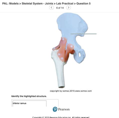
Table of Contents
PAL Models: Skeletal System Joints Quiz - A Comprehensive Guide
Understanding the skeletal system and its intricate network of joints is crucial for anyone studying anatomy, physiology, or related fields. This comprehensive guide delves deep into the world of skeletal joints, providing detailed explanations, interactive quiz questions, and valuable learning resources. We will explore the different classifications of joints, their structural components, and their functional roles within the human body. This guide is especially valuable for students using PAL (Personalized Assessment Learning) models, offering a robust framework for self-assessment and mastery of this important subject.
Understanding Joint Classification: A Foundation for Learning
Before diving into specific joint types, it's essential to grasp the basic classifications. Joints, also known as articulations, are points where two or more bones meet. They are categorized based on both their structural and functional characteristics:
Structural Classification: Focusing on Connective Tissue
This classification focuses on the type of connective tissue binding the bones:
-
Fibrous Joints: These joints are connected by fibrous connective tissue, offering little to no movement. Examples include sutures in the skull (immovable) and gomphoses (teeth in sockets). Think of the strong, inflexible nature of these connections.
-
Cartilaginous Joints: Characterized by cartilage connecting the bones, these joints allow for limited movement. Examples include synchondroses (hyaline cartilage, like the epiphyseal plates in growing bones) and symphyses (fibrocartilage, like the pubic symphysis). These joints offer a degree of flexibility and shock absorption.
-
Synovial Joints: These are the most common type of joint, characterized by a fluid-filled synovial cavity that allows for free movement. The presence of a synovial membrane, articular cartilage, and often ligaments contributes to their remarkable range of motion. We'll explore these further in the next section.
Functional Classification: Focusing on Range of Motion
This classification is based on the degree of movement the joint allows:
-
Synarthroses (Immovable): These joints have very little or no movement. Examples include sutures in the skull and gomphoses.
-
Amphiarthroses (Slightly Movable): These joints permit slight movement. Examples include intervertebral discs and the pubic symphysis.
-
Diarthroses (Freely Movable): These joints allow for a wide range of motion. All synovial joints fall under this category.
Synovial Joints: The Freely Movable Marvels
Synovial joints are the most versatile and complex type of joint in the body. Their structure facilitates a wide range of movement, crucial for activities of daily living. Let's examine their key components:
-
Articular Cartilage: A smooth layer of hyaline cartilage covering the articulating surfaces of the bones. It reduces friction and absorbs shock during movement. Think of it as nature's built-in shock absorber.
-
Synovial Cavity: A fluid-filled space between the articulating bones. This cavity allows for smooth movement.
-
Synovial Membrane: A thin membrane lining the synovial cavity. It secretes synovial fluid.
-
Synovial Fluid: A viscous fluid that lubricates the joint, reducing friction and providing nourishment to the articular cartilage. It's crucial for maintaining joint health and mobility.
-
Joint Capsule: A fibrous sac that encloses the synovial cavity, reinforcing the joint and providing stability.
-
Ligaments: Strong, fibrous bands of connective tissue that connect bones and reinforce the joint capsule. They provide stability and prevent excessive movement. Think of them as the joint's natural restraints.
-
Menisci (in some joints): Crescent-shaped pads of fibrocartilage that enhance joint stability and shock absorption. Found in the knee, they improve the congruency of the articulating surfaces.
-
Bursae (in some joints): Fluid-filled sacs located between the joint capsule and other structures like tendons or skin. They act as cushions and reduce friction.
Types of Synovial Joints: A Diverse Range of Motion
Synovial joints are further classified based on their shape and type of movement:
-
Plane Joints (Gliding Joints): These joints allow for gliding or sliding movements. Examples include the intercarpal and intertarsal joints. Think of the subtle movements of the wrist and ankle bones.
-
Hinge Joints: These joints allow for movement in one plane, like a door hinge. Examples include the elbow and knee joints. Consider the flexion and extension of your elbow when you bend your arm.
-
Pivot Joints: These joints allow for rotation around a single axis. Examples include the atlantoaxial joint (between the first and second cervical vertebrae) and the radioulnar joint (allowing for pronation and supination of the forearm). Imagine the turning motion of your head.
-
Condyloid Joints (Ellipsoid Joints): These joints allow for movement in two planes (flexion/extension and abduction/adduction). Examples include the radiocarpal joint (wrist) and metacarpophalangeal joints (knuckles). These joints enable a combination of movements.
-
Saddle Joints: These joints allow for movement in two planes, similar to condyloid joints, but with a greater range of motion. An example is the carpometacarpal joint of the thumb. The thumb's unique dexterity is due to this specialized joint.
-
Ball-and-Socket Joints: These joints allow for movement in three planes (flexion/extension, abduction/adduction, and rotation). Examples include the shoulder and hip joints. This joint type provides the greatest range of motion in the body.
PAL Models and Interactive Quizzes: Strengthening Your Understanding
Personalized Assessment Learning (PAL) models provide a powerful tool for mastering complex anatomical concepts. This approach emphasizes self-assessment, feedback, and iterative learning to solidify your understanding. To aid in your learning journey, here is an interactive quiz focusing on the different types of joints:
Quiz Questions:
-
Which type of joint is characterized by the presence of a synovial cavity? a) Fibrous joint b) Cartilaginous joint c) Synovial joint d) Gomphosis
-
The joint between the atlas and axis vertebrae is an example of which type of synovial joint? a) Hinge joint b) Pivot joint c) Saddle joint d) Ball-and-socket joint
-
Which type of joint allows for gliding movements? a) Hinge joint b) Condyloid joint c) Plane joint d) Pivot joint
-
What is the primary function of articular cartilage? a) Secretion of synovial fluid b) Reduction of friction and shock absorption c) Connection of bones d) Reinforcement of the joint capsule
-
Which of the following is NOT a component of a synovial joint? a) Synovial membrane b) Articular cartilage c) Symphysis d) Joint capsule
-
The knee joint is an example of which type of synovial joint? a) Hinge joint b) Condyloid joint c) Saddle joint d) Plane joint
Answer Key:
- c) Synovial joint
- b) Pivot joint
- c) Plane joint
- b) Reduction of friction and shock absorption
- c) Symphysis
- a) Hinge joint
This quiz serves as a starting point. Further quizzes can be developed to target specific aspects of joint structure, function, and associated pathologies. Remember to consult your textbook and other learning resources to enhance your understanding.
Beyond the Basics: Exploring Joint Pathology and Clinical Relevance
Understanding the structure and function of joints is fundamental to understanding various musculoskeletal disorders. Many conditions can affect joint health, impacting mobility and overall well-being. Here are a few examples:
-
Osteoarthritis: This degenerative joint disease is characterized by the breakdown of articular cartilage, leading to pain, stiffness, and reduced range of motion. It commonly affects weight-bearing joints like the knees and hips.
-
Rheumatoid Arthritis: This autoimmune disease causes inflammation of the synovial membrane, resulting in joint pain, swelling, and stiffness. It can affect multiple joints throughout the body.
-
Gout: This inflammatory condition is caused by the build-up of uric acid crystals in the joints, causing severe pain, swelling, and redness. It commonly affects the big toe.
-
Joint Injuries: Injuries such as sprains (ligament injuries), dislocations (displacement of bones from their normal position), and fractures (bone breaks) can significantly impair joint function.
Conclusion: Mastering Joint Anatomy Through PAL and Beyond
This comprehensive guide has explored the intricacies of the skeletal system's joints, encompassing their classification, structure, function, and clinical relevance. The integrated quiz provides a practical tool for self-assessment, aligned with the principles of Personalized Assessment Learning (PAL) models. By actively engaging with the material and using various learning strategies, you can build a solid foundation in this crucial area of anatomy. Remember that continuous learning and review are essential for long-term retention and mastery of complex anatomical concepts. Further research into specific joint pathologies and clinical applications will further enhance your understanding and prepare you for future studies in the field. Good luck!
Latest Posts
Latest Posts
-
Cold War Events And Policies Worksheet Answers
Mar 10, 2025
-
Ap Statistics Transformations To Achieve Linearity Worksheet
Mar 10, 2025
-
Match The Link State To The Interface And Protocol Status
Mar 10, 2025
-
Heming Way State Fall Camp Primitive
Mar 10, 2025
-
Which Pair Of Numbered Statements Best Completes
Mar 10, 2025
Related Post
Thank you for visiting our website which covers about Pal Models Skeletal System Joints Quiz . We hope the information provided has been useful to you. Feel free to contact us if you have any questions or need further assistance. See you next time and don't miss to bookmark.
