____ Posterior Film Placements Are Used In The Paralleling Technique.
Onlines
Apr 01, 2025 · 6 min read

Table of Contents
Posterior Film Placements in the Paralleling Technique: A Comprehensive Guide
The paralleling technique, a cornerstone of dental radiography, aims to produce high-quality, distortion-free radiographic images. Achieving this requires meticulous attention to detail, especially regarding film placement. While anterior film placement is relatively straightforward, posterior film placement presents unique challenges due to the anatomical complexities of the maxilla and mandible. This article delves into the intricacies of posterior film placements within the paralleling technique, offering a comprehensive guide for dental professionals seeking to master this crucial aspect of radiographic imaging.
Understanding the Paralleling Technique
Before exploring posterior film placements, let's revisit the fundamental principles of the paralleling technique. This technique utilizes a device, typically a long-cone paralleling instrument (Rinn XCP or similar), to maintain parallelism between the x-ray film and the long axis of the teeth being imaged. This parallelism minimizes image magnification and distortion, resulting in more accurate diagnostic images. The key to success lies in proper film positioning and beam alignment. Improper placement can lead to image elongation or foreshortening, hindering diagnostic accuracy.
Posterior Film Placement Challenges
The posterior region of the mouth presents several challenges for precise film placement:
-
Anatomical Obstructions: The presence of the tongue, palate, and alveolar ridges requires careful maneuvering of the film packet to ensure proper positioning without causing discomfort to the patient. The curved anatomy of the palate, in particular, demands a nuanced approach to maintaining parallelism.
-
Limited Space: The relatively limited space available in the posterior region necessitates precise placement to avoid overlapping structures and ensure that all the desired teeth are captured within the image. Overlapping teeth obscure diagnostic details, potentially masking important pathological findings.
-
Patient Comfort: Achieving the necessary parallelism while maintaining patient comfort is crucial. Improper positioning can cause discomfort or gagging, jeopardizing the quality of the radiograph and potentially leading to patient anxiety.
Maxillary Posterior Film Placement
Positioning films in the maxillary posterior region demands particular attention due to the palatal vault's curvature.
Step-by-step procedure:
-
Patient positioning: The patient should be seated comfortably with their head upright and positioned to allow for easy access to the posterior region of the mouth. Head positioning significantly impacts the accuracy of the radiograph.
-
Film selection: The appropriate size film (typically size 2 or 4) should be selected to encompass the desired teeth.
-
Film placement: The film is positioned in the mouth, aligning the long axis of the film parallel to the long axis of the maxillary posterior teeth. The film should be centered within the arch, ensuring all the relevant teeth are included within the image. The bite-wing tabs, if present, help ensure proper placement. The film should be close to the teeth, but not touching the palatal mucosa to avoid interference and image distortion.
-
Beam alignment: The x-ray beam is directed perpendicular to the film and the long axis of the teeth. The Rinn XCP instrument or similar device is vital here, maintaining this parallelism and stabilizing the film during exposure. Improper alignment will lead to distortion and inaccurate measurements.
-
Exposure: The x-ray exposure is made according to the manufacturer's instructions for the specific machine and film type. Overexposure or underexposure can both compromise the diagnostic quality of the image.
-
Image assessment: Once the exposure is complete, the image is assessed for proper exposure, alignment, and anatomical completeness. The radiograph should exhibit clear detail of the crowns and apices of all the maxillary posterior teeth.
Mandibular Posterior Film Placement
Mandibular posterior film placement presents its own set of challenges, primarily due to the tongue's presence and the proximity of the floor of the mouth.
Step-by-step procedure:
-
Patient positioning: Similar to maxillary positioning, ensure comfortable and upright head positioning for optimal image quality.
-
Film selection: Use appropriate sized films (size 2 or 4, depending on the area being imaged) to accurately capture the required teeth.
-
Film placement: This requires precise placement of the film against the lingual surfaces of the mandibular posterior teeth, while avoiding contact with the tongue. The film is positioned parallel to the long axis of the teeth. Patients may need guidance to position their tongue appropriately.
-
Beam alignment: As with maxillary placements, the Rinn XCP instrument or similar is essential for accurate beam alignment, ensuring perpendicularity to both the film and the long axes of the teeth.
-
Exposure: The x-ray exposure is made following manufacturer's instructions for the specific film and machine.
-
Image assessment: Upon image development, evaluate for optimal exposure, proper alignment, and complete representation of all relevant mandibular posterior teeth. Check for any signs of elongation or foreshortening.
Troubleshooting Common Errors in Posterior Film Placement
Several common errors can occur during posterior film placement. Recognizing and correcting these errors is crucial for producing diagnostic-quality radiographs.
-
Film bending or folding: Improper handling of the film can lead to bending or folding, resulting in artifacts on the radiograph. Careful handling is paramount.
-
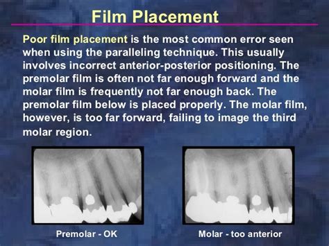
Film placement too far from the teeth: This results in magnification and distortion of the images. Maintain close proximity of the film to the lingual surfaces of the teeth, while avoiding contact with the soft tissues.
-
Incorrect beam alignment: This is a major cause of image distortion, leading to elongation or foreshortening. Use of the paralleling instrument is essential here.
-
Patient movement: Any movement by the patient during the exposure will blur the image and compromise diagnostic quality. Instructions for remaining still are crucial for patient cooperation.
-
Overlapping teeth: Poor film placement can lead to overlapping teeth, obscuring important details. Careful centering and precise placement are essential to avoid this.
-
Gagging reflex: This is a common issue with posterior film placements. Employ techniques like slow, deliberate film placement, distraction, and possibly using topical anesthetic agents to mitigate the gagging reflex.
Advanced Considerations
Beyond the basic techniques, several advanced considerations can improve the accuracy and consistency of posterior film placements.
-
Utilizing different types of paralleling instruments: Different instruments offer varying features and benefits. Consider exploring different instruments to find the one that suits your needs and preferences.
-
Digital radiography: Digital radiography offers numerous advantages, including reduced radiation exposure, immediate image viewing, and the ability to enhance or manipulate the image for better visualization. Digital systems should always be used following manufacturer's instructions for proper positioning and exposure techniques.
-
Understanding the anatomy of the region: A strong understanding of the anatomy of the maxilla and mandible is essential for proper film placement and interpretation of the resulting images. This knowledge enables anticipating potential challenges and finding solutions proactively.
-
Continuous training and practice: Proficiency in posterior film placement requires consistent training and practice. Regular review of the technique and hands-on practice will improve skill and ensure the production of high-quality images consistently.
Conclusion
Mastering posterior film placements within the paralleling technique is critical for obtaining high-quality, diagnostic-quality radiographs. Attention to detail in patient positioning, film placement, beam alignment, and image assessment are crucial aspects of the process. By understanding the unique challenges presented by the posterior region and employing the techniques and troubleshooting strategies outlined in this article, dental professionals can achieve a higher level of accuracy and consistency in their radiographic imaging, ultimately leading to improved patient care and diagnostic confidence. Continuous learning and refined techniques ensure that optimal image quality is achieved consistently, leading to better dental care. Remember that patient comfort and safety are paramount alongside achieving technically excellent radiographs.
Latest Posts
Latest Posts
-
Researchers Believe That Mental Recuperation Takes Place During
Apr 03, 2025
-
Ap Physics 1 Unit 7 Progress Check Mcq Part B
Apr 03, 2025
-
Summary Of Chapter 8 Of Animal Farm
Apr 03, 2025
-
Assisted Living Can Be Thought Of As A Combination Of
Apr 03, 2025
-
12 1 Identifying The Substance Of Genes
Apr 03, 2025
Related Post
Thank you for visiting our website which covers about ____ Posterior Film Placements Are Used In The Paralleling Technique. . We hope the information provided has been useful to you. Feel free to contact us if you have any questions or need further assistance. See you next time and don't miss to bookmark.
