Skeletal Muscle Concept Overview Physiology Interactive
Onlines
Mar 28, 2025 · 6 min read
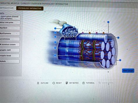
Table of Contents
Skeletal Muscle: A Concept Overview, Physiology, and Interactive Exploration
Skeletal muscle, the type of muscle tissue responsible for voluntary movement, is a fascinating and complex system. Understanding its structure, function, and physiology is crucial for anyone interested in exercise science, physical therapy, athletic performance, or simply maintaining a healthy lifestyle. This comprehensive article will delve into the intricacies of skeletal muscle, providing an overview of its concept, a detailed exploration of its physiology, and interactive elements to aid understanding.
I. A Conceptual Overview of Skeletal Muscle
Skeletal muscle, also known as striated muscle, is characterized by its striated appearance under a microscope, due to the highly organized arrangement of its contractile proteins. Unlike smooth muscle (found in internal organs) or cardiac muscle (found in the heart), skeletal muscle is under conscious control, allowing for precise and deliberate movements. It is responsible for a wide range of actions, from the subtle movements of the fingers to the powerful contractions involved in sprinting or weightlifting.
Key Characteristics:
- Voluntary Control: We consciously decide when and how to contract skeletal muscles.
- Striated Appearance: The organized arrangement of actin and myosin filaments creates a striped pattern.
- Multinucleated Cells: Each muscle fiber (cell) contains multiple nuclei.
- Attached to Bones: Most skeletal muscles are connected to bones via tendons, facilitating movement at joints.
- Rapid Contraction: Skeletal muscle contracts and relaxes relatively quickly.
- High Fatigue Rate: Sustained high-intensity activity can lead to muscle fatigue.
II. Delving into Skeletal Muscle Physiology: The Mechanism of Contraction
The ability of skeletal muscle to contract is driven by a complex interplay of cellular structures and biochemical processes. The process, known as the sliding filament theory, explains how muscle fibers shorten and generate force.
A. The Structural Components:
- Muscle Fiber (Myofiber): The individual muscle cell, containing many myofibrils.
- Myofibrils: Long cylindrical structures running the length of the muscle fiber, composed of repeating units called sarcomeres.
- Sarcomeres: The basic contractile unit of a muscle fiber, containing thick (myosin) and thin (actin) filaments.
- Myosin: Thick filaments with "heads" that bind to actin.
- Actin: Thin filaments with associated proteins (troponin and tropomyosin) regulating myosin binding.
- Sarcoplasmic Reticulum (SR): A specialized endoplasmic reticulum that stores calcium ions (Ca²⁺), essential for muscle contraction.
- Transverse Tubules (T-tubules): Invaginations of the sarcolemma (muscle cell membrane) that carry action potentials deep into the muscle fiber, ensuring uniform contraction.
B. The Excitation-Contraction Coupling:
- Nerve Impulse: A nerve impulse arrives at the neuromuscular junction, the point of contact between a motor neuron and a muscle fiber.
- Acetylcholine Release: The nerve impulse triggers the release of acetylcholine (ACh), a neurotransmitter.
- Sarcolemma Depolarization: ACh binds to receptors on the sarcolemma, causing depolarization – a change in the membrane potential.
- Action Potential Propagation: The action potential travels along the sarcolemma and down the T-tubules.
- Calcium Ion Release: The action potential triggers the release of Ca²⁺ from the SR into the sarcoplasm (cytoplasm of the muscle fiber).
- Cross-Bridge Cycling: Ca²⁺ binds to troponin, causing a conformational change that moves tropomyosin, exposing myosin-binding sites on actin. Myosin heads bind to actin, forming cross-bridges.
- Power Stroke: The myosin heads pivot, pulling the actin filaments towards the center of the sarcomere, causing muscle shortening.
- ATP Binding and Detachment: ATP binds to myosin, causing it to detach from actin.
- ATP Hydrolysis: ATP hydrolysis provides energy for the myosin head to return to its original position, ready for another cycle.
- Calcium Ion Replenishment: Ca²⁺ is actively pumped back into the SR, ending the contraction.
C. Types of Muscle Contractions:
- Isometric Contraction: Muscle length remains constant while tension increases (e.g., holding a weight).
- Isotonic Contraction: Muscle tension remains constant while muscle length changes (e.g., lifting a weight). This can be further divided into concentric (muscle shortens) and eccentric (muscle lengthens) contractions.
III. Energy Sources for Muscle Contraction:
Muscle contraction requires a significant amount of energy. This energy is primarily derived from the breakdown of ATP. However, the body utilizes various mechanisms to ensure a continuous supply of ATP for sustained muscle activity.
- Creatine Phosphate: A high-energy phosphate compound that quickly transfers its phosphate group to ADP, generating ATP.
- Anaerobic Glycolysis: The breakdown of glucose in the absence of oxygen, producing a small amount of ATP and lactic acid.
- Aerobic Respiration: The breakdown of glucose, fatty acids, and other fuels in the presence of oxygen, producing a large amount of ATP. This is the most efficient energy-producing pathway.
IV. Muscle Fiber Types:
Skeletal muscle fibers are not all created equal. They are classified into different types based on their contractile properties and metabolic characteristics.
- Type I (Slow-twitch): These fibers contract slowly, generate less force, but are highly resistant to fatigue. They rely primarily on aerobic respiration.
- Type IIa (Fast-twitch oxidative): These fibers contract faster, generate more force than Type I fibers, and have moderate fatigue resistance. They utilize both aerobic and anaerobic metabolism.
- Type IIx (Fast-twitch glycolytic): These fibers contract rapidly, generate the most force, but fatigue quickly. They primarily rely on anaerobic glycolysis.
The proportion of different fiber types varies between individuals and is influenced by genetics and training.
V. Interactive Exploration: A Simplified Model
(Note: A true interactive model would require a dynamic, web-based application. This section provides a textual representation to illustrate the concepts.)
Imagine a sarcomere represented by brackets [ ]. Within these brackets, we have actin filaments (represented by 'a') and myosin filaments (represented by 'm').
Initial State: [ aaaaaaaaa mmmmmmmm aaaaaaaaa ]
Calcium Ion Release (Ca²⁺): Ca²⁺ is released from the SR.
Cross-Bridge Cycling (Simplified):
- Binding: Myosin heads bind to actin: [ aaaaaammmmmma aaaaa ]
- Power Stroke: Myosin heads pivot, pulling actin filaments inwards: [ aaaaammmmmmaa aaaa ]
- Detachment and Reset: Myosin heads detach and reset: [ aaamm m mmmaa aa ] (Repeated cycles further shorten the sarcomere)
This simplified model helps visualize the sliding filament mechanism. A complete, interactive model would allow users to manipulate variables (Ca²⁺ concentration, ATP availability) and observe the effects on muscle contraction.
VI. Factors Affecting Muscle Function:
Several factors can influence skeletal muscle function, including:
- Age: Muscle mass and strength naturally decline with age (sarcopenia).
- Nutrition: Adequate protein intake is crucial for muscle growth and repair.
- Hormones: Hormones like testosterone and growth hormone play significant roles in muscle development.
- Exercise: Regular exercise, particularly resistance training, is essential for maintaining and increasing muscle mass and strength.
- Neuromuscular Coordination: The efficiency of communication between the nervous system and muscles greatly impacts performance.
- Muscle Fatigue: The inability of a muscle to maintain its force of contraction. This can be due to depletion of energy stores, accumulation of metabolic byproducts, or changes in neuromuscular transmission.
VII. Clinical Significance:
Understanding skeletal muscle physiology is crucial for diagnosing and treating various musculoskeletal disorders. These include:
- Muscle Dystrophies: Genetic disorders characterized by progressive muscle weakness and degeneration.
- Muscular Atrophy: Wasting away of muscle tissue, often due to disuse or nerve damage.
- Muscle Strains: Injuries caused by overstretching or tearing of muscle fibers.
- Fibromyalgia: A chronic condition characterized by widespread musculoskeletal pain.
VIII. Conclusion:
Skeletal muscle is a vital component of the human body, enabling movement, maintaining posture, and contributing to overall health and well-being. This article has provided a comprehensive overview of its structure, function, and physiology. Understanding these intricate mechanisms can help individuals optimize their fitness routines, prevent injuries, and appreciate the remarkable complexity of the human body. Further exploration through interactive resources and additional research will solidify understanding and encourage a deeper appreciation for the fascinating world of skeletal muscle.
Latest Posts
Latest Posts
-
Chapter 8 Summary Of The Hobbit
Mar 31, 2025
-
Elige El Preterito O El Imperfecto Para Completar La Historia
Mar 31, 2025
-
How Are Ip Headers Valuable For Security Analysts During Investigations
Mar 31, 2025
-
In All Furnace Cabinet Configurations The Return Air
Mar 31, 2025
-
Summary Of A Wall Of Fire Rising
Mar 31, 2025
Related Post
Thank you for visiting our website which covers about Skeletal Muscle Concept Overview Physiology Interactive . We hope the information provided has been useful to you. Feel free to contact us if you have any questions or need further assistance. See you next time and don't miss to bookmark.
