Which Bone-forming Process Is Shown In The Figure
Onlines
Mar 14, 2025 · 7 min read
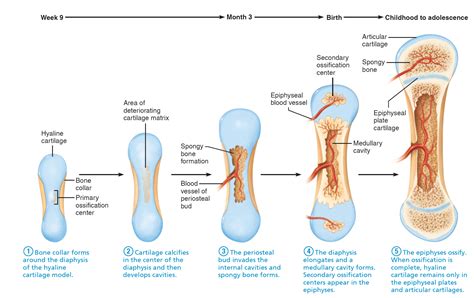
Table of Contents
Deciphering Bone Formation: Identifying the Process Depicted
The image (which is unfortunately not provided, as I am a text-based AI and cannot process images) likely depicts one of two primary bone-forming processes: intramembranous ossification or endochondral ossification. Understanding the key differences between these processes is crucial for identifying which one is shown in your figure. This article will delve into the details of each process, highlighting their distinct characteristics to aid you in your analysis. We'll explore the cellular mechanisms, the temporal sequence of events, and the resulting bone structures, providing a comprehensive framework for accurate identification.
Intramembranous Ossification: Direct Bone Formation
Intramembranous ossification, also known as direct bone formation, is a simpler process that forms the flat bones of the skull, the mandible, and the clavicles. It's characterized by the direct differentiation of mesenchymal cells into osteoblasts, the cells responsible for building bone. There's no cartilage intermediary involved.
Here's a breakdown of the key steps:
-
Mesenchymal Condensation: The process begins with the condensation of mesenchymal cells, a type of embryonic connective tissue. These cells cluster together at the site of future bone formation. This is a crucial first step, signaling the commitment to bone development.
-
Osteoblast Differentiation: Within the mesenchymal condensation, some cells differentiate into osteoblasts. Osteoblasts are specialized bone-forming cells that secrete osteoid, a specialized extracellular matrix consisting mainly of type I collagen. This osteoid provides the scaffold for the mineralization process.
-
Osteoid Secretion and Mineralization: Osteoblasts actively deposit osteoid, gradually building up a network of bony trabeculae (small, interconnected struts of bone). Simultaneously, the osteoid undergoes mineralization, a process where calcium phosphate crystals deposit within the matrix, hardening it into bone.
-
Trabecular Formation and Remodeling: As more osteoid is secreted and mineralized, the trabecular network expands and thickens. The spaces between the trabeculae are filled with hematopoietic tissue (blood cell-forming tissue), giving rise to the spongy bone structure characteristic of intramembranous ossification. Continuous bone remodeling – resorption and deposition – refines the bone structure, ensuring its strength and adaptation to mechanical stresses.
-
Formation of Periosteum: A fibrous membrane, the periosteum, forms around the developing bone. The periosteum plays a vital role in bone growth, repair, and nutrient supply.
Microscopic features to look for in an image depicting intramembranous ossification:
- Absence of cartilage: A key distinguishing feature is the lack of any cartilage template.
- Direct osteoblast differentiation from mesenchymal cells: You should be able to observe mesenchymal cells transforming directly into osteoblasts.
- Presence of woven bone: Initially, the bone formed is woven bone, characterized by a haphazard arrangement of collagen fibers. This is later remodeled into lamellar bone, which exhibits a more organized structure.
- Intramembranous bone formation within connective tissue: The process will be observed within a connective tissue environment, without the presence of a cartilage model.
Endochondral Ossification: Bone Formation via Cartilage Template
Endochondral ossification, also known as indirect bone formation, is a more complex process responsible for the formation of most of the bones in the body, particularly long bones. Unlike intramembranous ossification, this method uses a cartilage template as a precursor to bone formation.
Here's a detailed breakdown of the stages:
-
Cartilage Model Formation: The process begins with the formation of a hyaline cartilage model, a miniature version of the future bone. Mesenchymal cells differentiate into chondrocytes, which produce the cartilage matrix. This cartilage model serves as a scaffold for subsequent bone formation.
-
Bone Collar Formation: A collar of bone begins to form around the diaphysis (shaft) of the cartilage model. This collar is formed through intramembranous ossification, as osteoblasts from the surrounding perichondrium (cartilage's equivalent of periosteum) differentiate and deposit osteoid.
-
Primary Ossification Center Formation: Within the diaphysis, chondrocytes begin to hypertrophy (enlarge), leading to the degradation of the cartilage matrix. Blood vessels invade the hypertrophic cartilage, bringing in osteogenic cells (precursors to osteoblasts and osteoclasts) and nutrients. These osteoblasts then deposit bone matrix on the remnants of the cartilage, establishing the primary ossification center.
-
Secondary Ossification Center Formation: Similar processes occur in the epiphyses (ends) of the long bones, creating secondary ossification centers. However, the timing of their appearance varies depending on the bone.
-
Longitudinal Bone Growth: Growth in length occurs at the epiphyseal plates (growth plates), located between the epiphysis and metaphysis. Chondrocytes continue to proliferate and differentiate at these plates, pushing the epiphysis further away from the diaphysis. Simultaneously, bone is deposited behind the advancing cartilage, extending the length of the bone.
-
Bone Remodeling and Maturation: Throughout the entire process, continuous bone remodeling occurs, refining the bone structure, ensuring its strength, and adapting it to mechanical loading. The cartilage eventually disappears, leaving behind a fully ossified bone.
Microscopic features to look for in an image depicting endochondral ossification:
- Presence of cartilage: The presence of hyaline cartilage is a definitive indicator of endochondral ossification.
- Hypertrophic chondrocytes: Observe the enlarged chondrocytes that characterize the stages of cartilage matrix degradation.
- Bone formation within a cartilage template: The bone will be seen forming within the confines of the initial cartilage model.
- Presence of primary and secondary ossification centers: Identify the distinct locations of bone formation, reflecting the different stages of the process.
- Epiphyseal plates: Look for the characteristic growth plates where longitudinal bone growth occurs.
Distinguishing Intramembranous from Endochondral Ossification
The key differentiator lies in the presence or absence of a cartilage template. If the image shows direct bone formation from mesenchymal cells without any intervening cartilage, it's intramembranous ossification. If the image depicts bone formation within a pre-existing cartilage model, with visible hypertrophic chondrocytes and a progression of cartilage replacement by bone, it's endochondral ossification.
Remember to look for the microscopic features detailed above. The presence of woven bone versus lamellar bone, the arrangement of collagen fibers, and the relationship between bone and surrounding tissues are all crucial clues for accurate identification.
Beyond the Basics: Factors Influencing Bone Formation
Several factors influence both intramembranous and endochondral ossification:
-
Genetic factors: Genes play a crucial role in regulating the expression of various proteins involved in bone formation, influencing cell differentiation, matrix synthesis, and mineralization. Mutations in these genes can lead to various skeletal disorders.
-
Hormonal influences: Hormones such as growth hormone, parathyroid hormone, and vitamin D are critical for regulating bone growth and remodeling. Imbalances in these hormones can significantly affect bone development and integrity.
-
Mechanical stress: Mechanical loading, such as weight-bearing and muscle activity, influences bone remodeling. Bone adapts to the stresses placed upon it, increasing bone mass and density in areas of high stress.
-
Nutrition: Adequate intake of calcium, phosphorus, vitamin D, and other essential nutrients is essential for proper bone mineralization and growth. Deficiencies can lead to weakened bones and impaired bone development.
-
Inflammation: Inflammation can interfere with bone formation and remodeling, potentially leading to impaired bone growth and increased risk of fractures.
Clinical Relevance: Understanding Bone Formation Disorders
A thorough understanding of bone formation processes is critical for diagnosing and treating various bone disorders. Disruptions in either intramembranous or endochondral ossification can lead to a wide range of conditions, including:
-
Osteogenesis imperfecta: A genetic disorder affecting collagen synthesis, leading to fragile bones and increased fracture risk.
-
Achondroplasia: A genetic disorder affecting endochondral ossification, resulting in dwarfism.
-
Cleidocranial dysplasia: A genetic disorder affecting intramembranous ossification, leading to abnormalities in skull and clavicle development.
-
Osteopetrosis: A group of rare disorders characterized by excessive bone density, resulting from impaired bone resorption.
-
Rickets and osteomalacia: These metabolic bone diseases are caused by vitamin D deficiency, leading to impaired mineralization and weakened bones.
By carefully examining the microscopic details of your image and considering the characteristic features outlined in this article, you should be able to confidently identify the bone-forming process depicted. Remember that a deep understanding of these processes is crucial not only for scientific inquiry but also for comprehending various skeletal disorders and their treatments. Careful observation and a systematic approach will help unravel the mysteries of bone formation illustrated in your figure.
Latest Posts
Latest Posts
-
Final Exam For Is 100 C
Mar 14, 2025
-
Amoeba Sisters Video Recap Pedigrees Answer Key
Mar 14, 2025
-
1 The Five Common Types Of Expressway Interchanges Are
Mar 14, 2025
-
What Is The Authors Viewpoint In This Excerpt
Mar 14, 2025
-
Po Box 115009 Carrollton Tx 75011
Mar 14, 2025
Related Post
Thank you for visiting our website which covers about Which Bone-forming Process Is Shown In The Figure . We hope the information provided has been useful to you. Feel free to contact us if you have any questions or need further assistance. See you next time and don't miss to bookmark.
