Correctly Label The Following Anatomical Features Of The Eye
Onlines
Mar 13, 2025 · 7 min read
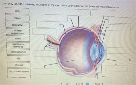
Table of Contents
Correctly Labeling the Anatomical Features of the Eye: A Comprehensive Guide
The human eye, a marvel of biological engineering, is a complex organ responsible for our sense of sight. Understanding its intricate anatomy is crucial for appreciating its function and diagnosing potential issues. This comprehensive guide will delve into the key anatomical features of the eye, providing detailed descriptions and assisting in their correct labeling. We'll cover everything from the outermost protective layers to the innermost light-sensitive structures. By the end, you'll be well-equipped to accurately identify and label the various components of this remarkable organ.
The Outermost Layer: Protection and Support
The outermost layer of the eye provides crucial protection and structural support. Its main components include:
1. Eyebrows (Supercilia):
These prominent ridges of hair above the eyes serve a vital protective function. They shield the eyes from sweat, rain, and other debris that could irritate the delicate surface. Their position and shape vary between individuals, contributing to facial recognition.
2. Eyelids (Palpebrae):
The upper and lower eyelids act as a protective shield, sweeping debris away from the cornea and lubricating the surface of the eye. Their rhythmic closure during blinking helps distribute tears, providing essential moisture and nourishment. The eyelids also contain glands that contribute to tear production, further enhancing their protective role. Meibomian glands, located within the eyelids, secrete an oily substance that prevents tear evaporation.
3. Eyelashes (Cilia):
These fine hairs lining the edges of the eyelids provide an additional layer of defense against foreign particles. Their sensitivity helps trigger the blink reflex, further protecting the cornea from damage. The eyelashes' angle and density vary considerably among individuals.
4. Conjunctiva:
A thin, transparent mucous membrane, the conjunctiva lines the inner surface of the eyelids (palpebral conjunctiva) and covers the sclera (bulbar conjunctiva). It keeps the eye moist and lubricated. Inflammation of the conjunctiva, known as conjunctivitis or pinkeye, is a common condition.
5. Lacrimal Apparatus:
This system comprises structures involved in tear production and drainage. The lacrimal gland, located in the upper outer corner of the eye socket, produces tears. Tears then drain through tiny canals (lacrimal canaliculi) into the lacrimal sac and subsequently into the nasolacrimal duct, which empties into the nasal cavity. Tears contain lysozyme, an enzyme that helps protect the eye from infection. Lacrimal puncta are the small openings where tears drain into the canaliculi.
The Middle Layer: The Vascular and Muscular Components
The middle layer of the eye, also known as the uvea, is rich in blood vessels and contains important muscular structures. Key components include:
6. Sclera:
The tough, white outer layer of the eye, the sclera, provides structural support and protection. It's the "white of the eye" visible around the iris. The sclera's fibrous nature helps maintain the eye's shape and protects its delicate inner structures.
7. Choroid:
Located beneath the sclera, the choroid is a highly vascular layer that nourishes the retina. Its rich blood supply is crucial for supplying oxygen and nutrients to the light-sensitive cells of the retina. The choroid's dark pigmentation helps absorb stray light, preventing internal reflections that could blur vision.
8. Ciliary Body:
This ring-shaped structure connects the choroid to the iris. The ciliary body contains the ciliary muscles, which control the shape of the lens, enabling accommodation – the eye's ability to focus on objects at varying distances. It also produces aqueous humor, the clear fluid filling the anterior chamber of the eye.
9. Iris:
The iris is the colored part of the eye, responsible for regulating the amount of light entering the eye. It contains two sets of muscles: the sphincter pupillae muscle, which constricts the pupil, and the dilator pupillae muscle, which dilates the pupil. The iris's color is determined by the amount and distribution of melanin.
10. Pupil:
The pupil is the black circular opening in the center of the iris. Its size adjusts based on the amount of ambient light, controlled by the iris muscles. In bright light, the pupil constricts to reduce the amount of light entering the eye, while in dim light, it dilates to maximize light intake.
The Innermost Layer: The Retina and Light Perception
The innermost layer of the eye, the retina, is responsible for converting light into electrical signals that are sent to the brain. This intricate structure contains millions of photoreceptor cells:
11. Retina:
The retina is a thin, light-sensitive layer lining the back of the eye. It contains millions of photoreceptor cells, including rods (responsible for vision in low light) and cones (responsible for color vision and visual acuity). The retina also contains specialized nerve cells that process visual information before sending it to the brain via the optic nerve.
12. Rods:
These photoreceptor cells are highly sensitive to light, enabling vision in low-light conditions. Rods are not responsible for color vision; they contribute mainly to peripheral vision and detecting movement.
13. Cones:
These photoreceptor cells are responsible for color vision and visual acuity (sharpness of vision). Cones are concentrated in the macula, particularly the fovea, which is the area of sharpest vision. There are three types of cones, each sensitive to a different range of wavelengths (red, green, and blue).
14. Macula:
This is a small, oval-shaped area in the center of the retina responsible for sharp, central vision. The macula contains a high concentration of cones, making it crucial for detailed vision. Age-related macular degeneration (AMD) is a common cause of vision loss affecting this area.
15. Fovea:
Located within the macula, the fovea is a small depression containing the highest concentration of cones. It's responsible for the sharpest, most detailed vision. The fovea is the point of clearest vision, used for focusing on fine details.
The Lens and the Refractive Media
The eye's optical system relies on several structures to focus light onto the retina:
16. Lens:
A transparent, biconvex structure located behind the iris, the lens plays a crucial role in focusing light onto the retina. The ciliary muscles change the lens's shape (accommodation), allowing the eye to focus on objects at different distances. With age, the lens loses flexibility, leading to presbyopia (age-related farsightedness).
17. Aqueous Humor:
This clear, watery fluid fills the anterior chamber of the eye (between the cornea and the lens). It helps maintain intraocular pressure and nourishes the cornea and lens. The production and drainage of aqueous humor are crucial for maintaining eye health. Blockage of drainage can lead to glaucoma.
18. Vitreous Humor:
A clear, gel-like substance that fills the posterior chamber of the eye (between the lens and the retina). The vitreous humor maintains the eye's shape and supports the retina. With age, it can shrink and detach, sometimes causing floaters or flashes of light.
19. Cornea:
The cornea is the transparent, dome-shaped outer layer of the eye, responsible for refracting (bending) light as it enters the eye. Its curved surface plays a significant role in focusing light onto the retina. The cornea is highly sensitive and is protected by the conjunctiva and eyelids.
Neural Pathways: Transmitting Visual Information
Once light is converted into electrical signals by the retina, this information needs to be transmitted to the brain:
20. Optic Nerve (II):
This cranial nerve carries visual information from the retina to the brain. The optic nerve consists of the axons of retinal ganglion cells, which transmit electrical signals generated by photoreceptors. The point where the optic nerve leaves the retina is known as the optic disc or blind spot, as it lacks photoreceptors.
21. Optic Chiasm:
At the optic chiasm, some nerve fibers from each optic nerve cross over to the opposite side of the brain. This crossing ensures that the right visual field is processed by the left side of the brain and vice versa. This allows for binocular vision and depth perception.
22. Optic Tract:
After the optic chiasm, the nerve fibers continue as the optic tract, carrying visual information to various brain regions for processing. The primary visual cortex in the occipital lobe is the main area for visual processing.
Conclusion: Mastering the Anatomy of the Eye
This detailed guide provides a comprehensive overview of the eye's anatomical features. Understanding these structures is essential for anyone studying ophthalmology, optometry, or related fields. Accurate labeling of these components requires a thorough grasp of their functions and interrelationships. By reviewing these components and their detailed descriptions, you will be well-prepared to correctly identify and label the numerous parts of the eye. Remember, this knowledge is not only academically valuable but also crucial for appreciating the complexity and delicate functionality of this remarkable sense organ. Further research into specific areas like retinal physiology or the intricacies of visual perception can provide even deeper understanding of this vital human organ.
Latest Posts
Latest Posts
-
General Ledger Questions Contain Multiple Tabs
Mar 13, 2025
-
Data Nuggets Deadly Windows Answer Key
Mar 13, 2025
-
What Problems Were The Gophers Causing On The Farm
Mar 13, 2025
-
Louisiana Class D Chauffeurs License Test
Mar 13, 2025
-
Practice Isotope Calculations 2 Answer Key
Mar 13, 2025
Related Post
Thank you for visiting our website which covers about Correctly Label The Following Anatomical Features Of The Eye . We hope the information provided has been useful to you. Feel free to contact us if you have any questions or need further assistance. See you next time and don't miss to bookmark.
