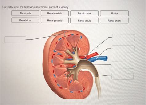
Correctly Label The Following Components Of The Kidney.
Onlines
Apr 02, 2025 · 7 min read

Table of Contents
Correctly Label the Following Components of the Kidney: A Comprehensive Guide
The kidney, a vital organ in the urinary system, plays a crucial role in maintaining homeostasis by filtering blood, removing waste products, and regulating fluid balance. Understanding its intricate structure is fundamental to appreciating its complex functions. This comprehensive guide will delve into the detailed anatomy of the kidney, enabling you to correctly label its various components. We'll explore each part, its function, and its relationship to the overall health of the urinary system.
Gross Anatomy of the Kidney: An Overview
Before diving into the microscopic details, let's establish a foundational understanding of the kidney's macroscopic features. Imagine a kidney as a bean-shaped organ, approximately the size of a fist. Each human typically possesses two kidneys, strategically located on either side of the vertebral column, nestled against the posterior abdominal wall. Several key external features characterize this organ:
1. Renal Capsule: The Protective Outer Layer
The renal capsule is a tough, fibrous membrane that encapsulates the entire kidney. This protective layer acts as the first line of defense against physical trauma and infection. Its smooth, glistening surface is essential for maintaining the kidney's shape and integrity.
2. Renal Cortex: The Outer Region
Beneath the renal capsule lies the renal cortex, a reddish-brown region that forms the outer layer of the kidney. This area is packed with nephrons, the functional units of the kidney responsible for filtering blood. The cortex has a granular appearance due to the densely packed glomeruli and convoluted tubules. It's where the initial stages of urine formation occur.
3. Renal Medulla: The Inner Region
The renal medulla is located deep to the cortex, characterized by its striated appearance. This region is comprised of cone-shaped structures called renal pyramids. These pyramids contain the loops of Henle and collecting ducts of the nephrons, crucial for concentrating urine. The medulla's striped pattern arises from the parallel arrangement of these structures.
4. Renal Columns: Extensions of the Cortex
Extending from the cortex into the medulla are the renal columns. These inward projections of cortical tissue separate adjacent renal pyramids. They provide structural support and vascular pathways connecting the cortex and medulla.
5. Renal Papilla: The Apex of the Pyramid
Each renal pyramid terminates in a pointed structure called the renal papilla. This papilla projects into a minor calyx, initiating the urine collection process. Multiple papillae drain into a single minor calyx.
6. Minor and Major Calyces: The Urine Collection System
The minor calyces are cup-like structures that surround the renal papillae, collecting urine. Several minor calyces converge to form major calyces. These major calyces then merge to create the renal pelvis.
7. Renal Pelvis: Funneling Urine to the Ureter
The renal pelvis is a funnel-shaped structure that acts as a reservoir for urine. It receives urine from the major calyces and channels it into the ureter, a tube that transports urine to the urinary bladder.
8. Hilum: The Entry and Exit Point
The hilum is a concave region on the medial border of the kidney where blood vessels, nerves, and the ureter enter and exit the kidney. This is a crucial area for the kidney's vascular supply and neural innervation.
9. Renal Artery and Vein: Blood Supply
The renal artery supplies oxygenated blood to the kidney, delivering a substantial volume of blood for filtration. The filtered blood, now depleted of waste products, exits the kidney via the renal vein. The extensive vascular network within the kidney is essential for its filtering function.
Microscopic Anatomy of the Kidney: The Nephron
The nephron, the functional unit of the kidney, is where the actual filtration and urine formation take place. Millions of nephrons are packed within the renal cortex and medulla, working in concert to maintain fluid and electrolyte balance. The nephron consists of several key structures:
1. Renal Corpuscle: The Filtration Unit
The renal corpuscle, also known as the Malpighian body, is the initial segment of the nephron. It consists of two main structures:
- Glomerulus: A network of capillaries where blood filtration occurs. The high pressure within the glomerulus forces fluid and small molecules (like water, glucose, amino acids, and waste products) out of the capillaries and into the Bowman's capsule.
- Bowman's Capsule (Glomerular Capsule): A double-walled cup-like structure that surrounds the glomerulus. It collects the filtrate from the glomerulus, initiating the urine formation process.
2. Renal Tubule: Modifying the Filtrate
The filtrate from Bowman's capsule enters the renal tubule, a long, convoluted structure where the composition of the filtrate is modified. The renal tubule consists of several segments:
- Proximal Convoluted Tubule (PCT): The initial segment of the renal tubule, characterized by its highly convoluted structure. It reabsorbs essential substances like glucose, amino acids, water, and electrolytes back into the bloodstream, while actively secreting certain substances into the filtrate.
- Loop of Henle: A U-shaped structure extending into the medulla. This loop plays a critical role in concentrating urine by establishing an osmotic gradient within the medulla. The descending limb is permeable to water, while the ascending limb is permeable to salts.
- Distal Convoluted Tubule (DCT): The final segment of the renal tubule before the collecting duct. It further modifies the filtrate by reabsorbing and secreting ions under hormonal regulation (e.g., aldosterone, antidiuretic hormone).
- Collecting Duct: This duct receives filtrate from multiple nephrons and plays a significant role in regulating water balance. The permeability of the collecting duct to water is controlled by antidiuretic hormone (ADH), influencing the final urine concentration.
The Juxtaglomerular Apparatus (JGA): Regulation of Blood Pressure
The juxtaglomerular apparatus (JGA) is a specialized structure located at the junction of the afferent arteriole and the distal convoluted tubule. It plays a crucial role in regulating blood pressure through the renin-angiotensin-aldosterone system (RAAS). The JGA comprises:
- Juxtaglomerular cells: Specialized smooth muscle cells in the afferent arteriole that secrete renin.
- Macula densa: A group of specialized epithelial cells in the distal convoluted tubule that detect changes in sodium concentration in the filtrate.
These components work together to monitor blood pressure and sodium levels, adjusting renin secretion accordingly to regulate blood volume and pressure.
Clinical Significance: Understanding Kidney Diseases
Understanding the kidney's anatomy is crucial for comprehending various kidney diseases and disorders. Disruptions in any of the structures discussed above can lead to significant health problems. For example:
- Glomerulonephritis: Inflammation of the glomeruli can impair their filtration function, leading to proteinuria (protein in the urine) and hematuria (blood in the urine).
- Kidney stones: These mineral deposits can form in the renal pelvis or calyces, causing pain and obstructing urine flow.
- Polycystic kidney disease (PKD): This genetic disorder results in the formation of cysts in the kidneys, reducing their function and potentially leading to kidney failure.
- Acute kidney injury (AKI): A sudden loss of kidney function, often caused by dehydration, infection, or medications.
- Chronic kidney disease (CKD): A gradual loss of kidney function over time, often caused by diabetes, high blood pressure, or glomerulonephritis.
Conclusion: Mastering the Anatomy of the Kidney
Correctly labeling the components of the kidney requires a thorough understanding of both its gross and microscopic anatomy. This comprehensive guide has provided a detailed overview of each structure, its function, and its clinical significance. By mastering the anatomical details, you will gain a deeper appreciation for the kidney's crucial role in maintaining human health and the implications of various kidney diseases. Remember, the intricate interplay between the different parts of the kidney ensures its efficient function in filtering blood, regulating fluid balance, and eliminating waste products. A clear understanding of this intricate system is vital for healthcare professionals and anyone interested in human physiology. Further study and visualization aids, such as anatomical models and diagrams, will solidify your understanding and enable you to accurately label every component of this vital organ.
Latest Posts
Latest Posts
-
Chapter 24 Pride And Prejudice Summary
Apr 03, 2025
-
6 14 Quiz New Threats And Responses
Apr 03, 2025
-
6 2 6 Install A Workstation Image Using Pxe
Apr 03, 2025
-
Prayers That Bring Healing By John Eckhardt Pdf
Apr 03, 2025
-
Radioactive Dating Game Lab Answer Key
Apr 03, 2025
Related Post
Thank you for visiting our website which covers about Correctly Label The Following Components Of The Kidney. . We hope the information provided has been useful to you. Feel free to contact us if you have any questions or need further assistance. See you next time and don't miss to bookmark.
