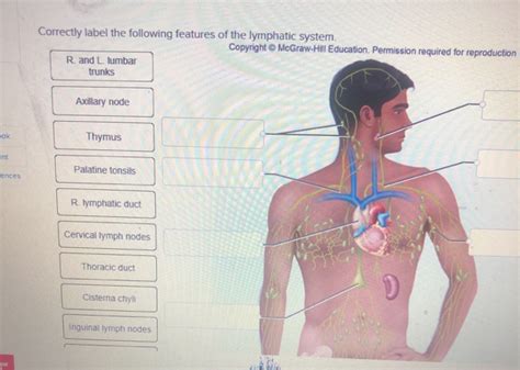
Correctly Label The Following Features Of The Lymphatic System
Onlines
Apr 05, 2025 · 6 min read

Table of Contents
Correctly Label the Following Features of the Lymphatic System: A Comprehensive Guide
The lymphatic system, often overlooked in discussions of the human body, plays a vital role in maintaining overall health and well-being. Understanding its intricate network of vessels, nodes, and organs is crucial for appreciating its functions in immunity, fluid balance, and lipid absorption. This comprehensive guide will delve into the key features of the lymphatic system, providing detailed descriptions and clarifying potential points of confusion. We'll equip you with the knowledge to correctly label its various components and understand their interconnected roles.
Major Components of the Lymphatic System: A Detailed Breakdown
The lymphatic system isn't a single, unified organ; rather, it's a complex network composed of several interconnected parts. Let's explore these crucial components:
1. Lymphatic Vessels: The Highways of the Lymphatic System
Imagine the lymphatic vessels as a network of one-way streets, carrying lymph – a clear, watery fluid – in a single direction toward the heart. These vessels are strategically distributed throughout the body, collecting interstitial fluid (fluid surrounding cells) and returning it to the bloodstream. The lymphatic vessels are categorized into several types:
-
Lymphatic Capillaries: These are the smallest lymphatic vessels, incredibly thin-walled and permeable, allowing interstitial fluid, proteins, and even larger particles (like bacteria) to enter. Their unique structure—with overlapping endothelial cells—prevents backflow.
-
Lymphatic Collecting Vessels: As lymphatic capillaries converge, they form larger collecting vessels. These vessels possess valves, ensuring unidirectional flow of lymph toward the lymph nodes. The smooth muscle within their walls helps propel the lymph forward.
-
Lymphatic Trunks: Multiple collecting vessels unite to form larger lymphatic trunks, each draining a specific region of the body. Examples include the lumbar, intestinal, bronchomediastinal, subclavian, and jugular trunks.
-
Lymphatic Ducts: Finally, the lymphatic trunks converge into two main lymphatic ducts: the right lymphatic duct and the thoracic duct. The right lymphatic duct drains lymph from the right upper quadrant of the body, while the thoracic duct, the larger of the two, drains lymph from the rest of the body. Both ducts empty into the venous system, returning the filtered lymph to the bloodstream.
Key Labeling Points: When labeling a diagram, clearly distinguish between the different vessel types based on their size and location within the lymphatic network. Remember to indicate the direction of lymph flow—always towards the heart.
2. Lymph Nodes: The Filters and Sentinels of the Immune System
Scattered along the lymphatic vessels are lymph nodes, small, bean-shaped structures acting as crucial filtering stations. These are not merely passive conduits; they are dynamic centers of immune activity. As lymph percolates through the lymph nodes, it encounters:
-
Lymphocytes: These immune cells—including T cells and B cells—actively monitor the lymph for foreign invaders like bacteria, viruses, and cancer cells. They mount an immune response, eliminating threats and preventing their spread.
-
Macrophages: These large phagocytic cells engulf and destroy pathogens and cellular debris within the lymph nodes. They play a crucial role in maintaining lymphatic fluid cleanliness.
Swollen Lymph Nodes (Lymphadenopathy): An increase in lymph node size often indicates an infection or other underlying condition. The body's immune system is working overtime, causing the nodes to swell as they combat the infection.
Key Labeling Points: Indicate the location of lymph nodes throughout the body, particularly in areas like the neck, armpits (axillae), groin (inguinal region), and mesentery. Highlight the afferent and efferent lymphatic vessels entering and exiting the nodes.
3. Lymphoid Organs: Specialized Centers of Immune Defense
Besides lymph nodes, the lymphatic system encompasses several specialized organs that contribute significantly to immune function:
-
Spleen: This fist-sized organ, located in the upper left abdomen, acts as a major filter for blood. It removes old or damaged red blood cells, recycles iron, and plays a crucial role in immune responses, housing lymphocytes and macrophages that monitor blood for pathogens.
-
Thymus: Located in the mediastinum (chest cavity), the thymus is particularly active during childhood and adolescence. It is the site where T lymphocytes mature and differentiate, becoming crucial components of the adaptive immune system. The thymus gradually atrophies with age.
-
Tonsils and Adenoids: These lymphoid tissues, located in the throat and nasopharynx, trap pathogens entering through the mouth and nose. They contribute to the body’s first line of defense against respiratory and oral infections.
-
Peyer's Patches: Located in the small intestine, these aggregates of lymphoid tissue monitor intestinal contents for pathogens. They play a vital role in gut immunity, preventing harmful bacteria from entering the bloodstream.
Key Labeling Points: Accurate placement of these organs is critical. Clearly distinguish the spleen from the liver and other abdominal organs. Show the location of the thymus in the mediastinum. Label the tonsils and adenoids in the throat area and Peyer's patches within the intestinal wall.
4. Lymph: The Fluid of the Lymphatic System
Lymph, the fluid transported by the lymphatic vessels, is essentially filtered interstitial fluid. It contains water, proteins, fats, and immune cells. Unlike blood, lymph lacks red blood cells. The composition of lymph varies depending on its location in the body, reflecting the local tissue environment. The movement of lymph, or lymphatic drainage, is facilitated by:
-
Skeletal Muscle Contractions: The rhythmic contractions of surrounding muscles compress lymphatic vessels, propelling lymph forward.
-
Respiratory Movements: The pressure changes during breathing assist in the movement of lymph.
-
Valves in Lymphatic Vessels: These prevent backflow, maintaining unidirectional lymph transport.
Key Labeling Points: While lymph itself isn't a structure to label visually, understanding its composition and movement is essential for a complete understanding of the lymphatic system.
Clinical Significance of the Lymphatic System
Disruptions in lymphatic function can lead to various health problems:
-
Lymphedema: This condition involves swelling due to impaired lymphatic drainage. It can be caused by infections, surgery, radiation therapy, or genetic factors.
-
Lymphoma: This is a type of cancer affecting the lymphatic system, originating in lymphocytes. It can manifest in various forms, depending on the type of lymphocyte involved.
-
Immunodeficiencies: Problems with the lymphatic system can impair the body's ability to fight infections.
Understanding the anatomy and function of the lymphatic system is critical for diagnosing and treating these conditions.
Interactive Exercises for Mastering Lymphatic System Anatomy
To reinforce your learning, try these exercises:
-
Labeling Diagram: Obtain a detailed diagram of the lymphatic system and label all the components discussed above. Pay close attention to the relationships between the different parts.
-
Tracing Lymph Flow: Trace the path of lymph from lymphatic capillaries to the venous system, highlighting the role of lymphatic vessels, lymph nodes, and lymphatic ducts.
-
Clinical Case Studies: Research case studies involving lymphatic system disorders (lymphedema, lymphoma). Analyze how disruptions in lymphatic function contribute to these conditions.
-
Compare and Contrast: Compare and contrast the lymphatic system with the cardiovascular system, highlighting their similarities and differences in structure and function.
Conclusion
The lymphatic system is a complex but crucial network responsible for maintaining fluid balance, lipid absorption, and immune defense. Understanding its intricate anatomy and physiology—from the smallest lymphatic capillaries to the largest lymphatic ducts and lymphoid organs—is fundamental to comprehending human health and disease. By mastering the correct labeling of its features, you'll gain a deeper appreciation for the vital role this often-overlooked system plays in our overall well-being. Regular review and application of the knowledge presented here will solidify your understanding and empower you to engage in more informed discussions about lymphatic health.
Latest Posts
Latest Posts
-
Summary Of I Am Legend Book
Apr 05, 2025
-
Accepting A Special Order Will Improve
Apr 05, 2025
-
Identify The Incorrect Statement Regarding The Vitreous Body
Apr 05, 2025
-
Which Of The Following Are Used To Provide Electric Heating
Apr 05, 2025
-
Activity Guide Input And Output Answer Key
Apr 05, 2025
Related Post
Thank you for visiting our website which covers about Correctly Label The Following Features Of The Lymphatic System . We hope the information provided has been useful to you. Feel free to contact us if you have any questions or need further assistance. See you next time and don't miss to bookmark.
