Correctly Label The Following Major Systemic Veins.
Onlines
Mar 18, 2025 · 6 min read
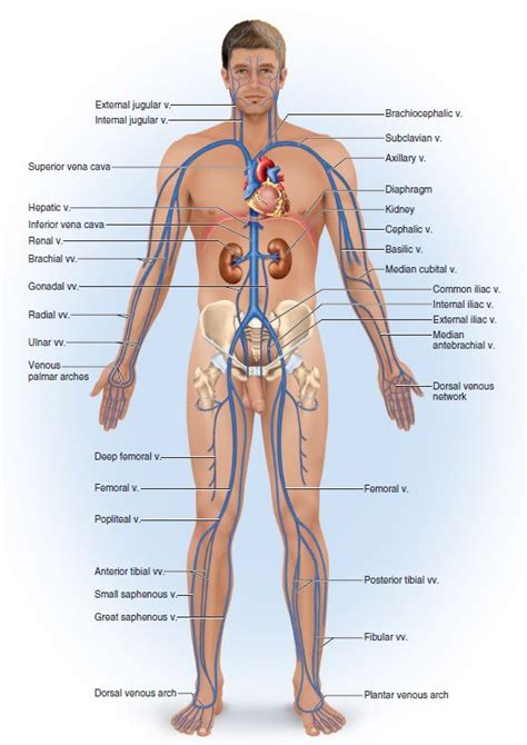
Table of Contents
Correctly Label the Following Major Systemic Veins: A Comprehensive Guide
The systemic venous system is a complex network responsible for returning deoxygenated blood from the body's tissues back to the heart. Understanding its intricate anatomy is crucial for medical professionals and students alike. This comprehensive guide will delve into the major systemic veins, providing detailed descriptions and aiding in their correct labeling. We'll explore their location, function, and clinical significance, ensuring a thorough understanding of this vital circulatory system.
The Superior Vena Cava and its Tributaries
The superior vena cava (SVC) is the large vein that receives deoxygenated blood from the upper body. It's a crucial component of the systemic venous system, playing a critical role in returning blood to the right atrium of the heart.
1. Brachiocephalic Veins:
The brachiocephalic veins are formed by the union of the internal jugular and subclavian veins on each side of the neck. They are significant because they act as the primary collecting veins for blood from the head, neck, and upper limbs.
-
Internal Jugular Veins: These veins drain blood from the brain, face, and neck. They are located alongside the internal carotid arteries. Their importance lies in their role in removing metabolic waste products and maintaining cerebral blood flow homeostasis. Damage to these veins can lead to significant complications.
-
Subclavian Veins: These veins collect blood from the upper limbs. They run beneath the clavicle (collarbone) and are essential for returning blood from the arms and shoulders to the heart. Their location makes them vulnerable to injury, particularly in cases of trauma.
2. Azygos Vein:
The azygos vein is a unique vein located on the right side of the vertebral column. It acts as a significant drainage pathway for the thoracic wall, and it receives blood from various smaller veins in the thorax. Its primary function is to collect blood that would otherwise be pooled in the posterior mediastinum. The azygos vein's anatomical variations are common and understanding these variations is crucial for accurate interpretation of medical imaging.
The Inferior Vena Cava and its Tributaries
The inferior vena cava (IVC) is the largest vein in the body. It receives deoxygenated blood from the lower body and empties into the right atrium of the heart. Understanding its tributaries is vital for comprehending the venous drainage of the lower extremities and abdominal viscera.
1. Common Iliac Veins:
The common iliac veins are formed by the union of the internal and external iliac veins on each side of the pelvis. They are essential for collecting blood from the lower limbs and pelvic organs. They then converge to form the inferior vena cava. Their location makes them susceptible to compression from surrounding structures.
-
External Iliac Veins: These veins drain blood from the lower limbs. They are located along the medial aspect of the thigh. Obstructions in these veins can lead to significant lower limb edema.
-
Internal Iliac Veins: These veins drain blood from the pelvic organs. They are located within the pelvic cavity and drain blood from the bladder, rectum, and uterus among other organs. Thrombosis in these veins can have serious consequences.
2. Hepatic Veins:
The hepatic veins are responsible for draining blood from the liver. They carry nutrient-rich blood from the liver's metabolic processes to the IVC. Understanding their function is crucial for assessing liver health and function. Impaired hepatic venous drainage can be indicative of various hepatic pathologies.
3. Renal Veins:
The renal veins drain blood from the kidneys. They are crucial for removing metabolic waste products and maintaining kidney homeostasis. Their location and close proximity to other major vessels make them susceptible to compression or injury. Abnormalities in renal venous drainage can indicate kidney disease or other systemic disorders.
4. Gonadal Veins:
The gonadal veins drain blood from the gonads (testes in males and ovaries in females). The right gonadal vein drains directly into the IVC, while the left gonadal vein drains into the left renal vein. These veins are important for transporting hormones and other substances produced by the gonads. Varicoceles (dilation of the pampiniform plexus) are a common condition affecting the gonadal veins in males.
5. Lumbar Veins:
The lumbar veins drain blood from the lumbar region of the back. They are important for collecting blood from the muscles and tissues of the lower back. These veins are often involved in the formation of the azygos and hemiazygos venous systems.
6. Phrenic Veins:
The phrenic veins drain blood from the diaphragm. They are essential for venous drainage of this crucial respiratory muscle. Their contribution to the IVC is relatively small compared to other tributaries.
Clinical Significance of Systemic Veins
Understanding the systemic venous system is not merely an academic exercise; it holds significant clinical relevance. Many diseases and conditions affect these veins, leading to various symptoms and complications.
1. Deep Vein Thrombosis (DVT):
DVT is the formation of a blood clot (thrombus) within a deep vein, often in the legs. This condition can be life-threatening if the clot travels to the lungs (pulmonary embolism). The location of the clot within the venous system directly influences its clinical significance.
2. Varicose Veins:
Varicose veins are enlarged, swollen, and twisted veins, most commonly seen in the legs. They are caused by incompetent venous valves, which allow blood to pool in the veins. This can result in discomfort, pain, and aesthetic concerns.
3. Venous Insufficiency:
Venous insufficiency is a condition where the veins are unable to effectively return blood to the heart. This can lead to edema, skin discoloration, and ulceration. Understanding the underlying anatomical pathways is crucial for diagnosis and management.
4. Superior Vena Cava Syndrome:
Superior vena cava syndrome occurs when the SVC is compressed or obstructed, usually due to a tumor. This can lead to facial edema, neck vein distention, and respiratory distress. Prompt diagnosis and treatment are vital to prevent serious complications.
Imaging Techniques for Visualizing Systemic Veins
Several imaging techniques are used to visualize and assess the systemic venous system. These techniques provide crucial information for diagnosing and managing various venous conditions.
1. Venography:
Venography is a technique where contrast material is injected into a vein, and X-rays are taken to visualize the vein's structure and patency. This is an invasive technique but provides highly detailed images.
2. Ultrasound:
Ultrasound is a non-invasive technique that uses sound waves to create images of the veins. It is commonly used to assess for DVT and other venous conditions.
3. CT and MRI:
CT (computed tomography) and MRI (magnetic resonance imaging) scans provide cross-sectional images of the body, which can be used to visualize the veins and surrounding structures. These techniques are particularly useful for assessing complex venous pathologies.
Conclusion
The systemic venous system is a complex yet crucial component of the circulatory system. Understanding the anatomy, function, and clinical significance of the major systemic veins is essential for healthcare professionals and students alike. This guide provides a comprehensive overview of this vital system, aiding in the correct labeling and understanding of its intricate network. Further exploration of specific veins and their associated pathologies will enhance your knowledge and contribute to improved patient care. Remember to always consult reputable anatomical resources and medical textbooks for the most accurate and up-to-date information.
Latest Posts
Latest Posts
-
Steady Precipitation Preceding A Front Is An Indication Of
Mar 19, 2025
-
Word 2021 In Practice Ch 2 Independent Project 2 4
Mar 19, 2025
-
When Inspecting The Brake Assembly You Should Ensure
Mar 19, 2025
-
Algebra Ii 5 2 Vertex Form Worksheet
Mar 19, 2025
-
Experiment 6 Acids Bases And Salts Report Sheet
Mar 19, 2025
Related Post
Thank you for visiting our website which covers about Correctly Label The Following Major Systemic Veins. . We hope the information provided has been useful to you. Feel free to contact us if you have any questions or need further assistance. See you next time and don't miss to bookmark.
