Label The Midsagittal Male Pelvis Using The Hints Provided.
Onlines
Mar 16, 2025 · 6 min read
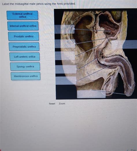
Table of Contents
Labeling the Midsagittal Male Pelvis: A Comprehensive Guide
The male pelvis, a complex structure crucial for weight bearing, locomotion, and protection of vital organs, presents a fascinating study in anatomy. Understanding its intricate components is fundamental to various fields, including medicine, physical therapy, and anthropology. This guide provides a detailed walkthrough of labeling a midsagittal view of the male pelvis, utilizing provided hints to enhance understanding and retention. We'll explore each bone, its features, and their functional significance.
The Bones of the Pelvis: A Deep Dive
The pelvic girdle, primarily responsible for transferring weight from the upper body to the lower limbs, is composed of three major bones: the two hip bones (ossa coxae, also known as innominate bones), and the sacrum. Let's examine each in detail within the context of a midsagittal view:
1. The Hip Bone (Os Coxae): A Tripartite Structure
The hip bone, a fusion of three distinct bones during development – the ilium, ischium, and pubis – presents a complex morphology. In a midsagittal view, the following features are particularly prominent:
-
Ilium: This large, flaring bone forms the superior portion of the hip bone. Its prominent features in a midsagittal section include:
- Iliac Crest: The superior, thickened border of the ilium, easily palpable externally. It serves as an important attachment site for numerous muscles.
- Anterior Superior Iliac Spine (ASIS): A prominent bony projection at the anterior end of the iliac crest. A crucial landmark for anatomical referencing and surgical procedures.
- Anterior Inferior Iliac Spine (AIIS): Located inferior to the ASIS, a less prominent but equally important feature.
- Posterior Superior Iliac Spine (PSIS): Located at the posterior end of the iliac crest, also important for anatomical reference.
- Posterior Inferior Iliac Spine (PIIS): Inferior to the PSIS.
- Arcuate Line: A curved line extending from the auricular surface of the ilium to the superior pubic ramus. It helps delineate the boundaries of the greater and lesser pelvis.
-
Ischium: This strong, posterior bone forms the inferoposterior part of the hip bone. Key features visible in the midsagittal section:
- Ischial Spine: A sharp projection located medially, marking the separation between the greater and lesser sciatic notches. Important for muscle attachment and ligamentous support.
- Ischial Tuberosity: A large, roughened projection forming the inferior portion of the ischium. It bears the weight of the body when sitting.
- Ischiopubic Ramus: Forms the inferior border of the obturator foramen.
-
Pubis: The anterior portion of the hip bone, articulated with its counterpart from the opposite hip bone at the pubic symphysis. Important features in a midsagittal section include:
- Superior Pubic Ramus: Extends superiorly from the pubic symphysis towards the acetabulum.
- Inferior Pubic Ramus: Extends inferiorly towards the ischium, helping to form the obturator foramen.
- Pubic Symphysis: A cartilaginous joint connecting the two pubic bones. Plays a significant role in childbirth by allowing some degree of movement.
- Pubic Crest: The thickened superior border of the pubic symphysis.
2. The Sacrum: The Keystone of the Pelvis
The sacrum, a triangular bone formed by the fusion of five sacral vertebrae, represents the central posterior element of the pelvic girdle. In a midsagittal section, its key features are:
- Sacral Promontory: The anterior projection of the superior border of the first sacral vertebra (S1). An important landmark for obstetrics and gynecology.
- Sacral Foramina: Paired openings on the anterior and posterior surfaces of the sacrum, providing passage for the sacral nerves. Only the anterior sacral foramina are visible in a midsagittal view.
- Sacral Canal: The continuation of the vertebral canal, housing the cauda equina. Clearly visible in the midsagittal plane.
- Sacral Hiatus: A gap at the inferior end of the sacral canal, where the sacral nerves exit.
- Apex of the Sacrum: The inferior, pointed end of the sacrum, articulating with the coccyx.
- Auricular Surface: The ear-shaped surface on the lateral aspect of the sacrum, articulating with the ilium to form the sacroiliac joint.
3. The Coccyx: A Vestige of the Tail
The coccyx, the vestigial remnant of the tail, is a small, triangular bone formed by the fusion of usually three to five coccygeal vertebrae. Its limited mobility is observed in a midsagittal view.
Functional Significance of Pelvic Features: Weight Bearing and Movement
The intricate anatomy of the male pelvis is directly related to its crucial functions. The large, robust structure of the hip bones and the sacrum provide a solid foundation for supporting the weight of the upper body. The shape and orientation of the pelvic bones also influence the gait and locomotion, enabling efficient bipedal movement.
The orientation of the acetabulum (the socket for the head of the femur), the strong ligaments surrounding the sacroiliac joints, and the sturdy muscles attached to the pelvic bones contribute to the stability and mobility of the pelvis. The strength of the ischial tuberosities is particularly relevant for supporting body weight during sitting. The shape of the pelvic inlet and outlet plays a crucial role in childbirth, although less prominent in a male pelvis.
The foramina in the sacrum and the obturator foramen in the hip bones are critical for the passage of nerves and blood vessels supplying the lower limbs and pelvic organs. Understanding the location of these openings is essential for various medical procedures.
Clinical Relevance: Fractures, Dislocations, and Other Conditions
The pelvic girdle, despite its robustness, is susceptible to various injuries and conditions:
- Pelvic Fractures: These can result from high-impact trauma and can involve any of the pelvic bones. The severity varies depending on the location and extent of the fracture.
- Sacroiliac Joint Dysfunction: This condition can cause pain and instability in the lower back and pelvis.
- Pubic Symphysis Diastasis: Separation of the pubic symphysis, often occurring during childbirth or due to trauma.
- Osteoarthritis: Degenerative joint disease affecting the sacroiliac and hip joints, causing pain and reduced mobility.
- Stress Fractures: Small cracks in the bone due to repetitive stress, such as running or other high-impact activities. More common in the ilium and pubic bones.
Labeling the Midsagittal Male Pelvis: A Step-by-Step Guide
Now, let's apply this knowledge to label a midsagittal view of the male pelvis. Refer to an anatomical image or diagram as you proceed through these steps. Remember to use precise anatomical terminology for accuracy.
-
Identify the major bones: Begin by identifying the ilium, ischium, and pubis of each hip bone, and the sacrum and coccyx.
-
Label the key features of the ilium: Mark the iliac crest, anterior superior iliac spine (ASIS), anterior inferior iliac spine (AIIS), posterior superior iliac spine (PSIS), posterior inferior iliac spine (PIIS), and the arcuate line.
-
Label the key features of the ischium: Locate and label the ischial spine and the ischial tuberosity.
-
Label the key features of the pubis: Identify and label the superior and inferior pubic rami, the pubic symphysis, and the pubic crest.
-
Label the key features of the sacrum: Mark the sacral promontory, anterior sacral foramina, the sacral canal, and the apex of the sacrum.
-
Label the coccyx: Identify and label the coccyx bone.
-
Verify your labels: Carefully check your labels to ensure accuracy and consistency with standard anatomical terminology.
By following these steps, you will build a strong understanding of the anatomy of the midsagittal male pelvis and its various components. Remember that consistent practice and review are key to mastering this complex subject. The more familiar you become with the bone structures and their relationships, the better you will be able to interpret anatomical images and apply this knowledge to various medical and clinical contexts. This detailed approach should enhance your understanding and ability to accurately label the midsagittal male pelvis. Continuous learning and reference to reputable anatomical resources are essential for strengthening your understanding.
Latest Posts
Latest Posts
-
One Source Of Lead On Some Job Sites Is
Mar 17, 2025
-
When Responding To Litigation Holds Foia Requests Investigations Or Inquiries
Mar 17, 2025
-
Participant Motivation Is Usually The Result Of
Mar 17, 2025
-
All Flags Such As Porn And Upsetting Offensive Are Query Independent
Mar 17, 2025
-
An Electrical Motor Provides 0 50 W Of Mechanical Power
Mar 17, 2025
Related Post
Thank you for visiting our website which covers about Label The Midsagittal Male Pelvis Using The Hints Provided. . We hope the information provided has been useful to you. Feel free to contact us if you have any questions or need further assistance. See you next time and don't miss to bookmark.
