Label The Structures Of The Pericardium In The Figure
Onlines
Apr 02, 2025 · 6 min read

Table of Contents
Labeling the Structures of the Pericardium: A Comprehensive Guide
The pericardium, a vital double-walled sac, encloses the heart and the roots of the great vessels. Understanding its intricate anatomy is crucial for comprehending cardiovascular physiology and pathology. This article provides a detailed guide to identifying and labeling the key structures of the pericardium, accompanied by visual aids and explanations to enhance your understanding.
The Pericardium: A Protective Sac
Before diving into the specific structures, let's establish a foundational understanding of the pericardium's role. This fibrous sac acts as a protective barrier, preventing the heart from overexpanding during periods of high blood volume, reducing friction during cardiac contractions, and providing structural support. Its multi-layered structure contributes to these crucial functions. We'll explore each layer in detail below.
Layers of the Pericardium: A Closer Look
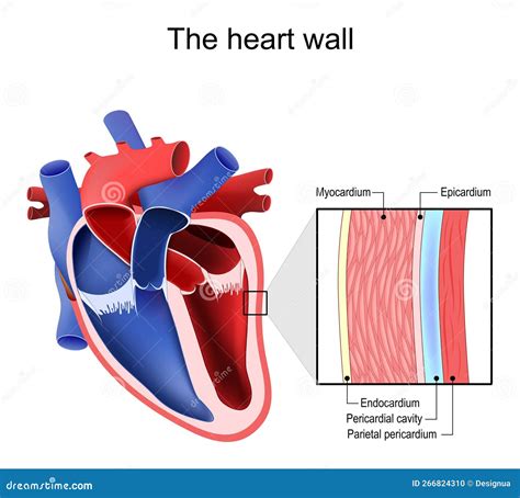
The pericardium is comprised of two main layers: the fibrous pericardium and the serous pericardium. The serous pericardium is further subdivided into two layers: the parietal layer and the visceral layer.
1. Fibrous Pericardium: The Outermost Layer
The fibrous pericardium, the outermost layer, is a tough, inelastic, dense connective tissue sac. Think of it as the strong, protective outer shell. Its primary function is to protect the heart from trauma and overdistension. It's this strong, rigid layer that prevents the heart from overstretching, even under significant pressure. It also anchors the heart to surrounding structures, preventing excessive movement within the thoracic cavity. This stability is crucial for optimal cardiac function. Key features to note are its fibrous nature and its relatively limited elasticity. Identifying this layer in a diagram is relatively straightforward due to its thickness and location.
2. Serous Pericardium: The Inner Protective Layer
Nestled within the fibrous pericardium lies the serous pericardium. This thinner, more delicate layer is responsible for creating a friction-free environment for the heart to beat within. It is made up of two continuous layers:
a) Parietal Pericardium: Lining the Fibrous Sac
The parietal pericardium is the outer layer of the serous pericardium, lining the inner surface of the fibrous pericardium. Think of it as the "wallpaper" of the fibrous pericardium's "room". It is a smooth, glistening membrane that adheres tightly to the fibrous pericardium. Its primary role is to create a smooth surface that minimizes friction between the heart and the surrounding structures during cardiac contractions.
b) Visceral Pericardium (Epicardium): Directly on the Heart
The visceral pericardium, also known as the epicardium, is the innermost layer of the serous pericardium. It's directly attached to the heart's surface and forms the outermost layer of the heart wall itself. It's intimately fused with the myocardium, the heart muscle. Its smooth surface minimizes friction during the rhythmic contractions and relaxations of the heart muscle. This layer plays a crucial role in lubricating the heart, ensuring efficient and less strenuous movements. It's vital to remember that the visceral pericardium and the epicardium are the same structure.
The Pericardial Cavity: A Space for Lubrication
Between the parietal and visceral layers of the serous pericardium lies the pericardial cavity. This is a potential space containing a small amount of serous fluid (approximately 15-50 ml). This fluid acts as a lubricant, significantly reducing friction between the heart and the pericardium during cardiac cycles. This friction reduction is essential for preventing damage and maintaining the efficiency of heart contractions. The lubrication in the pericardial cavity is crucial for allowing the heart to beat smoothly and efficiently without excessive wear and tear.
Clinical Significance: Pericardial Conditions
Understanding the pericardium's anatomy is critical in diagnosing and treating various clinical conditions. Problems within the pericardium can severely impact cardiac function.
Pericarditis: Inflammation of the Pericardium
Pericarditis, an inflammation of the pericardium, can cause chest pain, friction rubs (sounds of the inflamed layers rubbing against each other), and potentially life-threatening complications like cardiac tamponade. The cause can range from viral infections to autoimmune diseases or even post-surgical trauma.
Cardiac Tamponade: Life-Threatening Compression
Cardiac tamponade is a serious condition where fluid accumulation in the pericardial cavity compresses the heart, restricting its ability to fill with blood. This severely impairs cardiac output and can lead to circulatory collapse and death. Rapid diagnosis and intervention are crucial in these cases.
Pericardial Effusion: Fluid Buildup in the Pericardium
Pericardial effusion refers to an abnormal accumulation of fluid in the pericardial cavity. The fluid can be serous, hemorrhagic, or purulent depending on the underlying cause. While small effusions may be asymptomatic, large effusions can lead to cardiac tamponade.
Labeling the Structures in a Diagram: A Step-by-Step Guide
When labeling the structures of the pericardium in a diagram, follow these steps:
-
Identify the outermost layer: This is the fibrous pericardium. It's typically depicted as a thick, somewhat irregular layer surrounding the entire structure.
-
Locate the serous pericardium: This lies just beneath the fibrous pericardium.
-
Distinguish between the parietal and visceral layers: The parietal pericardium lines the inner surface of the fibrous pericardium, while the visceral pericardium (epicardium) directly covers the heart's surface. The space between these two layers is the pericardial cavity.
-
Label the pericardial cavity: This thin, fluid-filled space is crucial for minimizing friction during heart contractions.
-
Note the relationship between structures: Pay attention to how the layers are nested one within the other, and how the fluid-filled space contributes to efficient cardiac function.
Advanced Considerations: Connections and Clinical Relevance
The pericardium doesn't exist in isolation; it has several important anatomical connections. Understanding these connections provides a more holistic understanding of its role within the cardiovascular system. For example:
- Superior vena cava: The superior vena cava enters the right atrium, traversing close proximity to the pericardium.
- Inferior vena cava: Similar to the superior vena cava, its close proximity to the pericardium is important for contextual understanding.
- Pulmonary veins: The pulmonary veins enter the left atrium and run adjacent to the pericardium.
- Aorta and pulmonary artery: These great vessels leave the heart and are enveloped by the pericardium at their roots.
Understanding these connections helps interpret radiological images, grasp the implications of pericardial diseases, and perform accurate surgical interventions. For instance, during heart surgery, careful manipulation around the pericardium is crucial to avoid damage to surrounding structures and ensure a successful outcome.
Importance of Visual Aids: Diagrams and Images
Using diagrams and images is indispensable in mastering the anatomy of the pericardium. High-quality anatomical illustrations provide clear visual representations of the layers, their relationships, and the overall structure. Interactive online resources and anatomical atlases can be particularly useful in building a robust understanding.
Practicing labeling these structures on various diagrams strengthens your understanding and retention. The more you practice, the easier it will be to visualize and identify these components in real-world scenarios.
Conclusion: Mastering Pericardial Anatomy
Mastering the labeling of pericardial structures is crucial for anyone studying or working in the field of cardiovascular health. From the tough fibrous pericardium providing protection to the delicate serous pericardium ensuring smooth heart function, every layer plays a vital role. By understanding the layers, their relationships, and the clinical significance of pericardial conditions, you lay a solid foundation for a comprehensive understanding of cardiac anatomy and physiology. Remember, consistent practice using diagrams and a focus on the clinical implications will greatly enhance your learning and retention of this important anatomical structure.
Latest Posts
Latest Posts
-
The Given Reaction Proceeds In Two Parts
Apr 03, 2025
-
What Does A Facial Treatment Not Help To Relax
Apr 03, 2025
-
The Graph Represents The Hypothetical Market For Shrimp
Apr 03, 2025
-
What Is The Domain Of The Relation Graphed Below
Apr 03, 2025
-
Credit Card Utilization Penalty Points Are Charged Once Balance Exceeds
Apr 03, 2025
Related Post
Thank you for visiting our website which covers about Label The Structures Of The Pericardium In The Figure . We hope the information provided has been useful to you. Feel free to contact us if you have any questions or need further assistance. See you next time and don't miss to bookmark.
