Pal Histology Lymphatic System Lab Practical Question 1
Onlines
Mar 16, 2025 · 8 min read
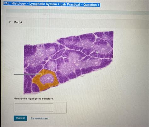
Table of Contents
Pal Histology: Lymphatic System Lab Practical - Question 1: A Deep Dive into Lymph Node Structure and Function
This article delves into the histological intricacies of the lymphatic system, specifically focusing on lymph nodes, a crucial component in immune response. We'll address common questions encountered in a histology lab practical, particularly those concerning lymph node structure and identification of key cellular components. Understanding the architecture of lymph nodes is paramount to comprehending their role in filtering lymph and initiating immune responses. This comprehensive guide will equip you with the knowledge to confidently tackle any lab practical questions related to lymph node histology.
Understanding the Lymphatic System's Role in Immunity
The lymphatic system acts as a crucial part of the body's defense system, working in conjunction with the immune system to protect against pathogens. It's a network of vessels, tissues, and organs that transport lymph, a fluid containing white blood cells and other immune cells. This fluid plays a key role in removing waste products, toxins, and pathogens from the body. Key components of this system include:
- Lymph: The fluid containing immune cells, waste products, and cellular debris.
- Lymphatic Vessels: A network of vessels that transport lymph throughout the body.
- Lymph Nodes: Bean-shaped organs that filter lymph and initiate immune responses.
- Spleen: Filters blood and plays a crucial role in immune response.
- Thymus: A gland that plays a critical role in the development of T lymphocytes (T cells).
- Tonsils and Adenoids: Collections of lymphoid tissue located in the throat and nasal passages.
Lymph Node Histology: A Microscopic View
Lymph nodes are highly organized structures with distinct regions, each performing a specific function. Understanding these regions is essential for proper identification in a histology lab setting. Let's examine the key features observable under a microscope:
1. Capsule and Trabeculae: The Structural Framework
The capsule, a dense connective tissue layer, encloses the lymph node, providing structural support. From this capsule, extensions called trabeculae project inward, dividing the node into compartments. These trabeculae serve as pathways for blood vessels and nerves to enter and exit the lymph node. Identifying the capsule and trabeculae is usually one of the first steps in analyzing a lymph node section. They're easily visible due to their dense collagenous nature.
2. Cortex: The Outer Region of Immune Activity
The cortex, the outer region of the lymph node, is characterized by several key features:
-
Cortical Nodules (Germinal Centers): These are highly organized areas within the cortex, often appearing as lighter staining regions. They are sites of B-lymphocyte proliferation and differentiation into plasma cells and memory B cells. Identifying germinal centers is crucial for confirming the presence of a lymph node. They are often surrounded by a darker staining mantle zone, rich in resting B cells. The size and presence of germinal centers can provide clues about the immune status of the individual.
-
Paracortex: Located between the cortex and medulla, the paracortex contains predominantly T lymphocytes, often appearing more densely packed than the surrounding areas. This area is also rich in high endothelial venules (HEVs), specialized postcapillary venules that allow the entry of lymphocytes from the blood into the lymph node. Recognizing HEVs can be challenging but is a valuable distinguishing feature. Their tall, cuboidal endothelial cells are distinct from the endothelial cells of other blood vessels.
3. Medulla: The Inner Region of Lymph Node
The medulla, the innermost region of the lymph node, is characterized by:
-
Medullary Cords: These are branching cords of lymphoid tissue containing a mixture of lymphocytes, macrophages, and plasma cells. They are generally less densely packed than the paracortex.
-
Medullary Sinuses: These are spaces between the medullary cords, filled with lymph and various immune cells. They're often visible as open spaces within the medulla. The medullary sinuses ultimately drain lymph into efferent lymphatic vessels.
4. Subcapsular Sinus: The Initial Filtering Point
The subcapsular sinus is a large, lymphatic sinus located directly beneath the capsule. It receives lymph from afferent lymphatic vessels. Macrophages within this sinus phagocytose antigens and other debris present in the lymph. This initial filtering step helps to remove a significant portion of foreign material before it reaches deeper regions of the lymph node.
5. Identifying Key Cell Types
Microscopic examination allows for the identification of various cell types within the lymph node. These include:
-
Lymphocytes: These are the predominant cell type, found throughout the lymph node in various regions. B lymphocytes are abundant in the cortical nodules, while T lymphocytes dominate the paracortex.
-
Macrophages: These large phagocytic cells are responsible for removing cellular debris and pathogens. They are found throughout the lymph node, particularly in the sinuses. Their large size and often foamy cytoplasm helps to distinguish them.
-
Plasma Cells: These antibody-producing cells are derived from B lymphocytes and are frequently observed in the medullary cords. Their characteristic eccentrically placed nucleus and abundant cytoplasm are useful identification features.
-
Dendritic Cells: These antigen-presenting cells play a critical role in initiating T cell responses. They are found throughout the lymph node, but are particularly abundant in the paracortex. Identification can be challenging and often requires specific staining techniques.
Common Lab Practical Questions and Answers
Here are some typical questions encountered in a histology lab practical focusing on lymph node structure:
Question 1: Identify the capsule, cortex, medulla, and trabeculae in this histological section of a lymph node. Describe their relative locations and appearances.
Answer: The capsule, a dense connective tissue layer, is the outermost structure. From the capsule, trabeculae, connective tissue extensions, project inward, dividing the node into compartments. The cortex is the outer region, characterized by the presence of cortical nodules (or germinal centers), light staining regions indicative of B-cell activity. The medulla, the inner region, consists of medullary cords and sinuses. The medullary cords are branching strands of lymphoid tissue, and the medullary sinuses are the spaces between the cords filled with lymph.
Question 2: Describe the cellular composition of the cortex and medulla. What are the functional implications of this cellular organization?
Answer: The cortex is predominantly composed of B lymphocytes concentrated in the cortical nodules (germinal centers), sites of B cell proliferation and antibody production. The paracortex (located between the cortex and medulla) is enriched with T lymphocytes. The medulla contains a mixture of lymphocytes, macrophages, and plasma cells arranged in medullary cords separated by medullary sinuses. This specific cellular organization reflects the functional roles of each region; the cortex is primarily involved in humoral immunity (antibody production), while the paracortex and medulla participate in cell-mediated immunity. The medullary sinuses facilitate the drainage of processed lymph.
Question 3: Identify the germinal centers within the cortex. Explain their significance in immune response.
Answer: Germinal centers are lighter staining areas within the cortex, often appearing as round or oval structures. They represent sites of intense B cell proliferation and differentiation. Within the germinal centers, B cells undergo somatic hypermutation, a process that improves the affinity of antibodies they produce. This selection of high-affinity B cells, coupled with the generation of memory B cells, is crucial for effective long-term immunity.
Question 4: Differentiate between afferent and efferent lymphatic vessels. Where would you expect to find them in relation to a lymph node?
Answer: Afferent lymphatic vessels carry lymph into the lymph node, while efferent lymphatic vessels carry lymph out of the lymph node. Afferent lymphatic vessels are found entering the lymph node at various points on its convex surface, while efferent lymphatic vessels exit the node at the hilum, a usually indented region on the concave side.
Question 5: What is the role of macrophages in lymph node function?
Answer: Macrophages are phagocytic cells that play a crucial role in removing cellular debris, pathogens, and other foreign material from the lymph. They reside in various locations within the lymph node, including the subcapsular sinus and medullary sinuses. Their phagocytic activity contributes to the overall filtering capacity of the lymph node. They also process antigens and present them to other immune cells, initiating further immune responses.
Advanced Considerations and Further Learning
Understanding lymph node histology goes beyond simply identifying structures. It involves understanding the intricate interplay of various cell types and their roles in the immune response. For a deeper understanding, consider exploring these advanced topics:
-
Immunohistochemistry: Techniques like immunostaining can help to specifically identify different cell types within the lymph node based on their unique protein expression.
-
Flow Cytometry: This technique allows for quantitative analysis of different cell populations within the lymph node, providing valuable insights into immune system dynamics.
-
Lymph Node Pathology: Studying the histological changes in lymph nodes associated with various diseases (e.g., infections, cancers) provides a deeper understanding of how the lymphatic system responds to pathological conditions.
By mastering the basic histological structures and cellular components of the lymph node, and exploring the advanced topics mentioned above, you'll build a strong foundation for understanding the crucial role of the lymphatic system in health and disease. This detailed understanding will prove invaluable, not only for acing your lab practical but for developing a strong grasp of immunology and related fields.
Latest Posts
Latest Posts
-
When Responding To Litigation Holds Foia Requests Investigations Or Inquiries
Mar 17, 2025
-
Participant Motivation Is Usually The Result Of
Mar 17, 2025
-
All Flags Such As Porn And Upsetting Offensive Are Query Independent
Mar 17, 2025
-
An Electrical Motor Provides 0 50 W Of Mechanical Power
Mar 17, 2025
-
Studying Marketing Should Help You To Blank
Mar 17, 2025
Related Post
Thank you for visiting our website which covers about Pal Histology Lymphatic System Lab Practical Question 1 . We hope the information provided has been useful to you. Feel free to contact us if you have any questions or need further assistance. See you next time and don't miss to bookmark.
