The Adult Hip Bone Consists Of _____ Regions.
Onlines
Apr 02, 2025 · 7 min read

Table of Contents
The Adult Hip Bone Consists of Three Regions: A Deep Dive into Pelvic Anatomy
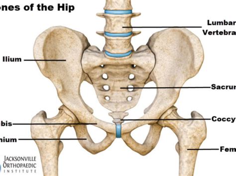
The adult hip bone, also known as the coxal bone or innominate bone, is a complex structure crucial for weight-bearing, locomotion, and protection of internal organs. Contrary to what some simplified diagrams might suggest, it's not a single, monolithic bone. Instead, the adult hip bone is formed by the fusion of three distinct bones: the ilium, the ischium, and the pubis. Understanding the anatomy of these three regions is fundamental to comprehending the biomechanics of the hip joint and diagnosing a wide array of pelvic pathologies. This article provides a comprehensive exploration of each region, their fusion points, and their clinical significance.
The Ilium: The Superior Wing of the Hip Bone
The ilium is the largest of the three hip bones, forming the superior and lateral portions of the hip. Its prominent features are easily palpable and contribute significantly to the overall shape and function of the pelvis.
Key Anatomical Features of the Ilium:
-
Iliac Crest: This is the superior border of the ilium, a thick, curved ridge that runs from the anterior superior iliac spine (ASIS) to the posterior superior iliac spine (PSIS). It serves as an important landmark for many clinical procedures and is a common site for muscle attachment. The iliac tubercle, a roughened area on the external lip of the iliac crest, is also a significant attachment point for muscles.
-
Anterior Superior Iliac Spine (ASIS): This is the most anterior projection of the iliac crest and serves as an important attachment site for muscles such as the sartorius and tensor fasciae latae. It's easily palpable and used as a reference point in many clinical examinations.
-
Anterior Inferior Iliac Spine (AIIS): Located inferior to the ASIS, the AIIS is a less prominent projection serving as an attachment point for the rectus femoris muscle.
-
Posterior Superior Iliac Spine (PSIS): The most posterior projection of the iliac crest, the PSIS is also an important landmark, notably serving as an attachment for the erector spinae muscles.
-
Posterior Inferior Iliac Spine (PIIS): Inferior to the PSIS, the PIIS is a less prominent spine.
-
Greater Sciatic Notch: A large, curved indentation on the posterior aspect of the ilium, it forms the superior boundary of the greater sciatic foramen. This foramen is crucial for the passage of numerous nerves and blood vessels.
-
Auricular Surface: This is a rough, articular surface on the medial aspect of the ilium that articulates with the sacrum, forming the sacroiliac joint (SIJ). This joint plays a critical role in transmitting weight from the upper body to the lower limbs.
-
Iliac Fossa: A concave surface on the internal aspect of the ilium, it provides extensive area for muscle attachment and protects the abdominal viscera.
The Ischium: The Inferior and Posterior Portion of the Hip Bone
The ischium forms the inferior and posterior portions of the hip bone. It is characterized by its strong and robust structure, reflecting its role in weight-bearing and providing support for the body in the seated position.
Key Anatomical Features of the Ischium:
-
Ischial Spine: A sharp projection that projects medially into the pelvic cavity. It serves as an important landmark and attachment point for ligaments and muscles.
-
Ischial Tuberosity: This is a large, roughened projection that forms the bony part of the buttock. It is the weight-bearing portion of the hip bone when sitting, making it crucial for postural stability. It is also an attachment point for numerous muscles of the thigh and hip.
-
Lesser Sciatic Notch: Located inferior to the ischial spine, the lesser sciatic notch contributes to the formation of the lesser sciatic foramen, another passageway for nerves and blood vessels.
-
Ischiopubic Ramus: This contributes to the formation of the obturator foramen, a large opening in the hip bone that's closed by a membrane except for small openings for nerves and blood vessels.
The Pubis: The Anterior and Inferior Portion of the Hip Bone
The pubis forms the anterior and inferior part of the hip bone. It is involved in the formation of the pubic symphysis, a joint that plays a significant role during pregnancy and childbirth.
Key Anatomical Features of the Pubis:
-
Superior Ramus: Extends posteriorly and superiorly towards the acetabulum.
-
Inferior Ramus: Extends inferiorly and laterally to meet with the ischial ramus.
-
Pubic Tubercle: A prominent elevation on the superior ramus, serving as an important landmark and muscle attachment site.
-
Pubic Symphysis: The joint where the two pubic bones articulate with each other, joined by a fibrocartilaginous disc. This joint allows for slight movement, which is particularly important during pregnancy and childbirth.
-
Obturator Foramen: The large opening formed by the ischiopubic rami, serving as a pathway for blood vessels and nerves.
The Acetabulum: The Hip Socket
The acetabulum is a cup-shaped structure formed by the fusion of the ilium, ischium, and pubis. It's the socket that receives the head of the femur, forming the hip joint. Its deep socket and strong surrounding ligaments contribute to the hip's remarkable stability and strength. Understanding the acetabulum is key to comprehending hip function and pathology, including hip dysplasia and fractures. The acetabular labrum, a fibrocartilaginous ring, deepens the acetabulum and enhances stability.
Fusion and Clinical Significance
The fusion of the ilium, ischium, and pubis occurs during adolescence, typically completed by the age of 16-18. The fusion points are crucial areas to consider in evaluating fractures and other pelvic injuries. Incomplete fusion (as seen in certain developmental disorders) can lead to instability and functional impairments.
Clinical Relevance: Fractures and Other Injuries
The hip bone's three regions are susceptible to different types of fractures depending on the mechanism of injury and the age of the individual.
-
Iliac Wing Fractures: Usually caused by high-energy trauma, these fractures often involve the body of the ilium and are often associated with other pelvic injuries.
-
Acetabular Fractures: These complex fractures involve the socket of the hip joint and often require surgical intervention. They are frequently associated with significant complications and rehabilitation needs.
-
Ischial Tuberosity Fractures: These fractures are commonly caused by falls or direct trauma to the buttocks, often affecting older adults with osteoporotic bones.
-
Pubic Rami Fractures: Usually caused by direct trauma or compression forces, these fractures may involve the superior or inferior pubic rami. They are often associated with urinary complications.
-
Sacroiliac Joint Dysfunction: Problems with the SIJ, such as inflammation or instability, can cause significant pain and disability, often requiring specialized treatment.
Imaging Techniques for Evaluating the Hip Bone
Several imaging techniques are used to visualize the hip bone and diagnose associated pathologies:
-
X-rays: Provide basic information about the bone structure and help identify fractures and other bony abnormalities.
-
CT scans: Offer detailed cross-sectional images of the bone, providing excellent visualization of complex fractures and other abnormalities.
-
MRI: Provides high-resolution images of both bone and soft tissues, useful in evaluating ligament injuries, muscle tears, and other soft tissue pathologies.
Conclusion
The adult hip bone is a complex and crucial structure composed of three distinct regions: the ilium, the ischium, and the pubis. Understanding the anatomy of each region, their fusion points, and their clinical significance is essential for healthcare professionals involved in the diagnosis and treatment of pelvic pathologies. The intricate interplay of bone, joint, and muscle creates a robust structure capable of supporting the body, facilitating movement, and protecting vital internal organs. A thorough understanding of its multifaceted anatomy is critical for anyone studying human anatomy, biomechanics, or the diagnosis and treatment of pelvic injuries. The robust nature of the hip bone reflects millions of years of evolutionary adaptation, making it a testament to the effectiveness of biological design. Further study into the subtleties of hip bone anatomy will continue to refine our understanding of this remarkable structure and improve patient care.
Latest Posts
Latest Posts
-
The Graph Represents The Hypothetical Market For Shrimp
Apr 03, 2025
-
What Is The Domain Of The Relation Graphed Below
Apr 03, 2025
-
Credit Card Utilization Penalty Points Are Charged Once Balance Exceeds
Apr 03, 2025
-
Maggie Plans A Workout For Tuesday
Apr 03, 2025
-
How Many Chapters Are In Crime And Punishment
Apr 03, 2025
Related Post
Thank you for visiting our website which covers about The Adult Hip Bone Consists Of _____ Regions. . We hope the information provided has been useful to you. Feel free to contact us if you have any questions or need further assistance. See you next time and don't miss to bookmark.
