Pal Cadaver Appendicular Skeleton Pectoral Girdle Lab Practical Question 1
Onlines
Mar 18, 2025 · 7 min read
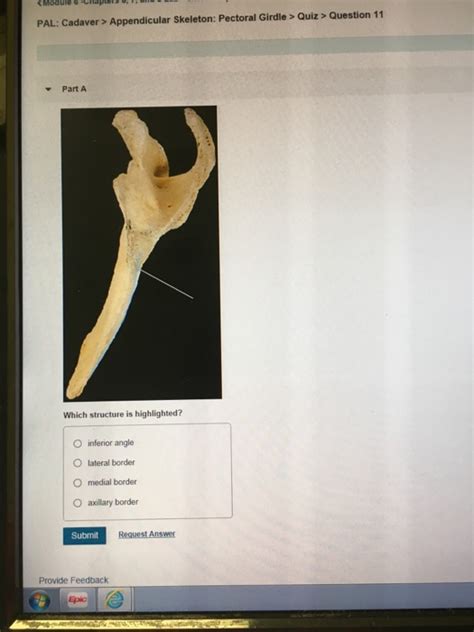
Table of Contents
Pal Cadaver Appendicular Skeleton: Pectoral Girdle - Lab Practical Question 1: A Comprehensive Guide
This article delves deep into the intricacies of the appendicular skeleton, specifically focusing on the pectoral girdle, within the context of a pal cadaver lab practical. We’ll explore the key anatomical structures, their functions, and common questions encountered in such practical examinations. This comprehensive guide is designed to equip students with the knowledge and understanding needed to confidently navigate this topic. We'll tackle "Lab Practical Question 1" by breaking down the likely questions and providing detailed answers.
Understanding the Appendicular Skeleton
The human skeleton is broadly divided into two main parts: the axial skeleton (skull, vertebral column, rib cage) and the appendicular skeleton. The appendicular skeleton comprises the bones of the limbs (upper and lower) and their respective girdles that connect them to the axial skeleton. The pectoral girdle, also known as the shoulder girdle, is the crucial link connecting the upper limbs to the axial skeleton. Its proper functioning is essential for arm movement and overall upper body mobility. A thorough understanding of its components is therefore critical for any anatomy student.
Components of the Pectoral Girdle
The pectoral girdle is primarily composed of two bones on each side of the body:
-
Clavicle (Collarbone): A long, S-shaped bone that lies horizontally across the upper chest. It articulates medially with the sternum (breastbone) at the sternoclavicular joint and laterally with the scapula (shoulder blade) at the acromioclavicular joint. Its primary function is to transmit forces from the upper limb to the axial skeleton, providing stability and preventing excessive movement of the shoulder. In lab practicals, you should be able to identify its distinct curves and articulating surfaces.
-
Scapula (Shoulder Blade): A flat, triangular bone located on the posterior aspect of the thorax. It possesses several important features crucial for muscle attachment and articulation:
- Acromion: A lateral projection forming the highest point of the shoulder. It articulates with the clavicle.
- Coracoid Process: A hook-like projection extending anteriorly. It provides attachment points for muscles of the shoulder and upper arm.
- Glenoid Cavity (Fossa): A shallow, concave surface on the lateral aspect of the scapula. This is where the head of the humerus (upper arm bone) articulates to form the glenohumeral joint (shoulder joint). Note the relatively small size of the glenoid cavity which contributes to the shoulder's considerable range of motion but also its inherent instability.
- Spine: A prominent ridge running across the posterior surface of the scapula, providing attachment sites for several back and shoulder muscles.
- Supraspinous Fossa and Infraspinous Fossa: Depressions superior and inferior to the spine, respectively, providing origins for important rotator cuff muscles.
- Subscapular Fossa: A large depression on the anterior surface of the scapula.
Articulations of the Pectoral Girdle
The pectoral girdle possesses several crucial articulations:
-
Sternoclavicular Joint (SC Joint): This is a saddle joint connecting the medial end of the clavicle to the sternum. It allows for a relatively wide range of motion, including elevation, depression, protraction, and retraction of the clavicle. Understanding the ligaments stabilizing this joint is crucial.
-
Acromioclavicular Joint (AC Joint): A plane joint connecting the acromion process of the scapula to the lateral end of the clavicle. It permits limited gliding movements between these bones.
-
Glenohumeral Joint (Shoulder Joint): While not strictly part of the pectoral girdle itself, it is intimately connected and crucial for understanding upper limb movement. This is a ball-and-socket joint, providing the widest range of motion of any joint in the body. Look for the glenoid labrum, a fibrocartilaginous ring that deepens the glenoid cavity and enhances stability.
Muscles Associated with the Pectoral Girdle
Numerous muscles originate or insert on the bones of the pectoral girdle, contributing to the complex movements of the shoulder and arm. Some key muscles to be familiar with for your practical include:
- Trapezius: A large, superficial muscle covering much of the upper back and neck, contributing to scapular elevation, depression, rotation, and retraction.
- Rhomboids (major and minor): Deep muscles located between the scapula and vertebral column, involved in scapular retraction and downward rotation.
- Levator Scapulae: Elevates the scapula.
- Pectoralis Minor: Depresses the scapula and protracts the shoulder.
- Serratus Anterior: Protracts the scapula and rotates it upward.
- Deltoids: A powerful muscle responsible for shoulder abduction, flexion, and extension. This is often a key muscle highlighted in lab practicals.
- Rotator Cuff Muscles (supraspinatus, infraspinatus, teres minor, subscapularis): These four muscles originate on the scapula and insert on the humerus. Their primary function is to stabilize the shoulder joint and assist with rotation. Damage to these muscles is a common clinical issue.
Lab Practical Question 1: Anticipated Scenarios
Now, let's address the anticipated questions within the context of "Lab Practical Question 1" focusing on the pal cadaver appendicular skeleton and pectoral girdle:
Scenario 1: Identification and Articulations
- Question: Identify the bones of the pectoral girdle and describe their articulations with other bones.
- Answer: This requires you to accurately identify the clavicle and scapula on the cadaver. You'll then need to precisely describe the sternoclavicular joint, acromioclavicular joint, and indirectly, the glenohumeral joint, highlighting the types of joints and the movements they allow. Mention relevant ligaments supporting these articulations (e.g., coracoclavicular, acromioclavicular, sternoclavicular ligaments). Demonstrate your understanding of the bony landmarks involved in each articulation.
Scenario 2: Muscle Attachments and Actions
- Question: Identify the origin and insertion points of at least three muscles that attach to the scapula and describe their actions.
- Answer: This requires you to locate the origins and insertions of muscles on the scapula, ideally on the actual cadaver. For example, you could choose the trapezius, deltoid, and rhomboids. You then need to clearly describe the specific actions of each muscle on the scapula and the shoulder joint. Use precise anatomical terminology to describe the movements (e.g., abduction, adduction, elevation, depression, protraction, retraction, medial rotation, lateral rotation).
Scenario 3: Clinical Significance
- Question: Discuss the clinical significance of a fracture of the clavicle.
- Answer: This question tests your understanding of the functional importance of the clavicle. You should discuss the potential complications of a clavicular fracture, such as pain, impaired shoulder movement, and potential damage to nearby nerves and blood vessels. Mention the typical mechanism of injury and the various treatment options. You could also discuss the importance of the clavicle in force transmission from the upper limb to the axial skeleton and how a fracture might compromise this.
Scenario 4: Comparative Anatomy
- Question: Compare and contrast the pectoral girdle of a human with that of another species (e.g., a cat or a bird).
- Answer: This question tests your broader understanding of the pectoral girdle and its adaptations across different species. You should highlight the similarities and differences in bone structure, articulations, and musculature between the human pectoral girdle and the chosen species. Explain how these differences reflect the varying locomotor requirements of the different animals. For example, a bird's pectoral girdle is adapted for flight with a keeled sternum for flight muscle attachment, whereas a human's is more adapted for dexterity and manipulation.
Scenario 5: Palpation and Identification
- Question: Locate and palpate the clavicle and acromion process on a living subject.
- Answer: Although this might not be directly on the cadaver, it is a realistic component of a practical exam. You'll need to demonstrate your ability to accurately identify these bony landmarks by touch on a human subject (this may be part of a simulated scenario using a model). Explain your methodology and any safety considerations.
Preparing for the Lab Practical
To excel in your lab practical, consider these preparation strategies:
- Thorough Study: Review anatomical diagrams, models, and textbooks. Focus on the detailed features of the clavicle and scapula.
- Hands-on Practice: If possible, spend time observing and handling real bones or anatomical models. This will help you familiarize yourself with the texture, shape, and size of the bones.
- Active Recall: Test yourself repeatedly using flashcards or practice questions. This helps solidify your understanding and improves retention.
- Work with Peers: Study with classmates and quiz each other. Teaching the material to others strengthens your own comprehension.
- Understand Function: Don't just memorize structures; understand their functions and how they work together. This provides a deeper level of understanding.
By following this detailed guide and practicing diligently, you will be well-prepared to confidently answer any question related to the pal cadaver appendicular skeleton, particularly concerning the pectoral girdle, during your lab practical. Remember to utilize the cadaver as a primary learning tool, actively exploring the anatomy and integrating your theoretical knowledge with practical observation. Good luck!
Latest Posts
Latest Posts
-
Each Of The Following Graphs Shows A Hypothetical Relationship
Mar 18, 2025
-
Experiment 10 Report Sheet Vinegar Analysis
Mar 18, 2025
-
Simulation Lab 13 2 Module 13 Configuring The User Account Control
Mar 18, 2025
-
Pal Cadaver Appendicular Skeleton Upper Limb Lab Practical Question 3
Mar 18, 2025
-
Which Of The Following Describes How Probe Data Is Collected
Mar 18, 2025
Related Post
Thank you for visiting our website which covers about Pal Cadaver Appendicular Skeleton Pectoral Girdle Lab Practical Question 1 . We hope the information provided has been useful to you. Feel free to contact us if you have any questions or need further assistance. See you next time and don't miss to bookmark.
